
Preparation method of soluble VP1 antigen of pig hidroa

A soluble, swine vesicular disease virus technology, applied in the fields of botanical equipment and methods, biochemical equipment and methods, chemical instruments and methods, etc., can solve problems such as reducing the formation of inclusion bodies, and achieve the effect of high purity

Inactive Publication Date: 2008-10-15
LANZHOU INST OF VETERINARY SCI CHINESE ACAD OF AGRI SCI
View PDF0 Cites 5 Cited by
  • Summary
  • Abstract
  • Description
  • Claims
  • Application Information

AI Technical Summary

Problems solved by technology

[0006] The technical problem to be solved in the present invention is to express and produce soluble VP1 protein with good biological activity in Escherichia coli, reduce the formation of inclusion bodies, and avoid complicated denaturation and renaturation processes

Method used


Image

  • Preparation method of soluble VP1 antigen of pig hidroa

Examples


Embodiment

[0034] The following examples are further descriptions of the present invention, which do not constitute any limitation to the scope of the present invention.

Embodiment 1

[0041] Pick a single colony of rVP1 and inoculate it in 3 mL of 2×YT medium containing 100 μg / mL ampicillin, culture it with shaking at 37°C for at least 16 hours, take 500 μL and inoculate it in 50 mL LB medium, and culture it at 37°C until the OD600 reaches 0.8~ 1.0, add IPTG to a final concentration of 0.10 mmol / mL, place the culture flask in a temperature-controlled shaker at 18°C, and incubate at 250 rpm for 16 hours. 1×PBS (pH7.3) was used to collect the bacteria, and the precipitate was collected after centrifugation. The bacteria were lysed by ultrasonication in an ice bath and centrifuged at high speed.

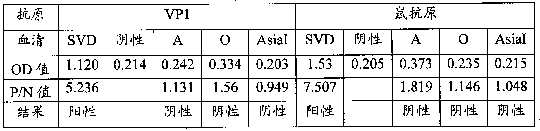
[0042] SDS-PAGE protein electrophoresis showed that the lysed bacterial supernatant had an obvious protein band at 19KDa; Western blotting showed that the band could be recognized by SVDV-specific positive serum.

[0043]Filter the supernatant after ultrasonically lysing the cells, transfer the filtrate to a chromatographic column prepacked with Ni-NTAHis Bind Resin,...

Embodiment 2

[0045] The expression of rVP1 was induced and the obtained VP1 protein was purified as in Example 1, except that the final concentration of IPTG was 0.05 mmol / mL, and the induction temperature was 16°C.

[0046] SDS-PAGE protein electrophoresis showed that the lysed bacterial supernatant had an obvious protein band at 19KDa; Western blotting showed that the band could be recognized by SVDV-specific positive serum.


PUM


Abstract

The invention discloses a method for producing soluble VP1 antigen for detection of swine vesicular disease virus, which comprises the following steps of: 1). cloning a VP1 gene; 2). constructing rVP1; 3). Inducing the expression of rVP1 to generate soluble VP1; and 4). Purifying the soluble VP1. An inducer is IPTG with a concentration of between 0.05 to 1.00 mmol / mL; the induction temperature is between 16 and 20 DEG C. The method generates soluble VP1 protein in colon bacilli, reducing the production of inclusion bodies, avoiding complex processes of denaturation and renaturation. The obtained VP1 protein is high in purity, the same as SVDV particles in terms of immune trait and can be used as an antigen to replace SVDV particles to establish an indirect detection method for sensitive, specific, safe and reliable SVDV serum antibody.

Description

technical field [0001] The invention relates to a preparation method of swine vesicular disease VP1 antigen, more specifically, a preparation method of soluble swine vesicular disease VP1 antigen. Background technique [0002] Swine Vesicular Disease (SVD) is an acute infectious disease of pigs caused by the Swine Vesicular Disease Virus (SVDV) of the family Picornaviridae Enterovirus. Blisters or festers are very similar to porcine foot-and-mouth disease and are easily misdiagnosed. Therefore, its prevention and treatment has attracted widespread attention. [0003] At present, porcine vesicular disease has been eradicated in my country, so the use of intact virus particles to establish a diagnostic method has the potential risk of spreading the virus, which is not advisable in practice. [0004] The SVDV genome contains a large open reading frame, encoding a polyprotein consisting of 2185 amino acids, in which the encoded structural proteins are VP4 (1A, 69aa), VP2 (1B, 2...

Claims


Application Information

Patent Timeline
IPC IPC(8): C12N15/41C12N15/70C07K14/085G01N33/569
Inventor 刘湘涛尹双辉尚佑军田宏孙世琪
Owner LANZHOU INST OF VETERINARY SCI CHINESE ACAD OF AGRI SCI
Learn More