Human primary tumor cell separation preparation method

A primary tumor cell and cell technology, applied in cell dissociation methods, tumor/cancer cells, biochemical equipment and methods, etc., can solve the problems of low separation rate and slow proliferation of human primary tumor cells

Inactive Publication Date: 2017-11-24
CENTURY BIOSTRENGTH BEIJING PTY LTD
View PDF2 Cites 2 Cited by
  • Summary
  • Abstract
  • Description
  • Claims
  • Application Information

AI Technical Summary

Problems solved by technology

[0003] Aiming at the above problems such as low separation rate and slow proliferation of human primary tumor cells, the present invention provides a method for separating and preparing human primary tumor cells. a lot of time

Method used


Image

  • Human primary tumor cell separation preparation method
  • Human primary tumor cell separation preparation method
  • Human primary tumor cell separation preparation method

Examples


Embodiment 1

[0019] A method for separating and preparing human primary tumor cells, characterized in that the method comprises the following steps:

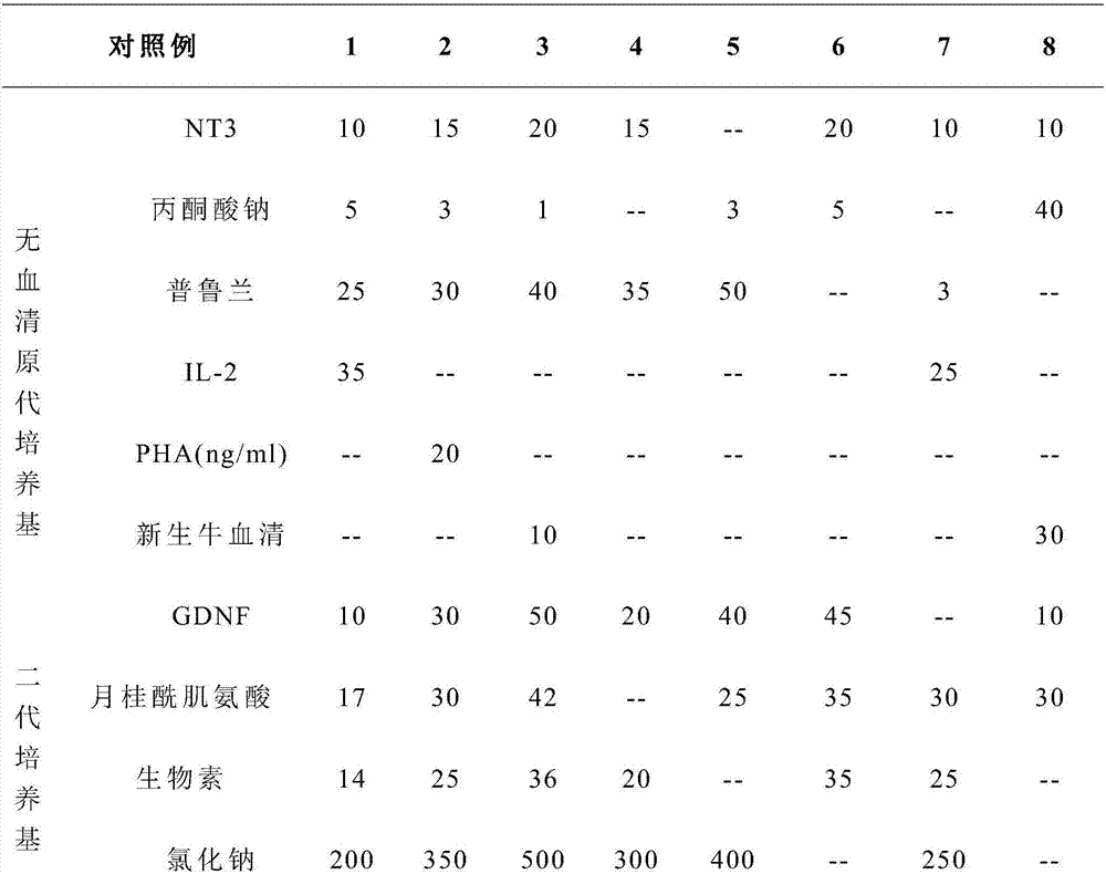
[0020] 1) Rinse the isolated tumor tissue with 0.9% sodium chloride injection, remove blood stains, cut off necrosis and connective tissue;

[0021] 2) Cut the tumor tissue obtained in step 1) into 1mm 3 Size, placed in a centrifuge tube, add 15ml 0.1% type I collagenase to the centrifuge tube, cold digest overnight at 4°C, add 5ml 0.1mg / ml hyaluronidase and 10ml 10μl / ml DNA the next day Enzyme, digested in a shaker at 40°C for 6 hours, passed the digested mixture through a 40 μm cell mesh, collected the filtrate, centrifuged for 5 minutes, and removed the supernatant to obtain a single cell suspension;

[0022] 3) Add serum-free primary medium to the single-cell suspension obtained in step 2), pipette and mix, count the cells, inoculate in culture bottles, add serum-free primary medium to each bottle, and place it in a carbon dioxide therm...

Embodiment 2

[0029] A method for separating and preparing human primary tumor cells, characterized in that the method comprises the following steps:

[0030] 1) Rinse the isolated tumor tissue with 0.9% sodium chloride injection, remove blood stains, cut off necrosis and connective tissue;

[0031] 2) Cut the tumor tissue obtained in step 1) into 1mm 3 Size, placed in a centrifuge tube, add 5ml 0.1% type I collagenase to the centrifuge tube, cold digest overnight at 4°C, add 1ml 0.1mg / ml hyaluronidase and 5ml 10μl / ml DNA the next day Enzyme, digested in a shaker at 34°C for 4 hours, passed the digested mixture through a 40 μm cell sieve, collected the filtrate, centrifuged for 1 min, and removed the supernatant to obtain a single cell suspension;

[0032] 3) Add serum-free primary medium to the single-cell suspension obtained in step 2), pipette and mix, count the cells, inoculate in culture bottles, add serum-free primary medium to each bottle, and place it in a carbon dioxide thermostat...

Embodiment 3

[0039] A method for separating and preparing human primary tumor cells, characterized in that the method comprises the following steps:

[0040] 1) Rinse the isolated tumor tissue with 0.9% sodium chloride injection, remove blood stains, cut off necrosis and connective tissue;

[0041] 2) Cut the tumor tissue obtained in step 1) into 1mm 3 Size, placed in a centrifuge tube, add 10ml 0.1% type I collagenase to the centrifuge tube, cold digest overnight at 4°C, add 3ml 0.1mg / ml hyaluronidase and 8ml 10μl / ml DNA the next day Enzyme, digested in a shaker at 36°C for 5 hours, passed the digested mixture through a 40 μm cell sieve, collected the filtrate, centrifuged for 3 minutes, and removed the supernatant to obtain a single cell suspension;

[0042] 3) Add serum-free primary medium to the single-cell suspension obtained in step 2), pipette and mix, count the cells, inoculate in culture bottles, add serum-free primary medium to each bottle, and place it in a carbon dioxide therm...


PUM


Abstract

The invention relates to a human primary tumor cell separation preparation method which comprises the following steps of washing tumor cells which are separated out by using a 0.9 percent sodium chloride injection, removing blood, and shearing necrotic and paeoniae tissues; shearing the tumor tissues into 1mm<3>, placing the shorn tumor tissues in a centrifugal tube, adding type I collagenase in the centrifugal tube, carrying out cold digestion overnight, adding hyaluronidase and DNA enzyme next day, carrying out digestion, sieving a mixed liquid after digestion by a cell screen, collecting a filtrate, and carrying out centrifugation to remove supernatant to obtain a single-cell suspension; adding a serum-free primary culture medium, beating, uniformly mixing, counting cells, inoculating the cells into a culture bottle, supplementing the serum-free primary culture medium, and placing the culture bottle in a carbon dioxide constant-temperature and constant-humidity incubator to carry out culture; and carrying out cryopreservation. The human primary tumor cell separation preparation method provided by the invention is high in harvest rate of prepared cells, enables the cells to be rapid in proliferation, can improve the proliferation capability of human primary tumor cells, can increase the proliferation speed of the human primary tumor cells, reduces the culture period and has prominent progress.

Description

technical field [0001] The invention relates to the field of tumor cells and primary culture of tumor cells, in particular to a method for separating and preparing human primary tumor cells. Background technique [0002] Tumor is a new organism formed when cells in local tissues lose control of their growth at the gene level under the action of various tumorigenic factors, resulting in abnormal proliferation of cells. With the improvement of people's material and cultural living standards, life behavior Due to the changes in the way of life, the increasingly serious air and environmental pollution, and the prolongation of the life expectancy of the aging population, the death spectrum of the population in China has undergone great changes. The main cause of death has become a social problem that people pay attention to. Tumor is one of the common malignant tumors, with the highest incidence rate in the age group of 40 to 50 years old. According to the world epidemiological s...

Claims


Application Information

Patent Timeline
Patent Type & Authority Applications(China)
IPC IPC(8): C12N5/09A01N1/02
CPCA01N1/0221C12N5/0693C12N2500/12C12N2500/90C12N2501/13C12N2501/90C12N2509/00
Inventor 李霞云潘新贺伟刘世红卢家堃
Owner CENTURY BIOSTRENGTH BEIJING PTY LTD
Why Eureka
Try Eureka