
A screening method for cervical precancerous lesions combining spectroscopy and images

A cervical cancer screening technology, applied in the field of spectral analysis and medical image processing, can solve the problems of low cost, low specificity, and underdeveloped medical resources.

Active Publication Date: 2022-04-08
NANJING UNIV OF AERONAUTICS & ASTRONAUTICS
View PDF0 Cites 1 Cited by
  • Summary
  • Abstract
  • Description
  • Claims
  • Application Information

AI Technical Summary

Problems solved by technology

This method is low in cost and can be applied at the grassroots level, but the specificity is not high
At present, there is still a lack of a cervical cancer screening method with good sensitivity and specificity and low price in clinical practice, which can be applied to primary hospitals with underdeveloped medical resources to improve the screening rate and accuracy of cervical cancer.

Method used


Image

  • A screening method for cervical precancerous lesions combining spectroscopy and images
  • A screening method for cervical precancerous lesions combining spectroscopy and images
  • A screening method for cervical precancerous lesions combining spectroscopy and images

Examples


Embodiment Construction

[0029] The invention will be described in further detail below in conjunction with the accompanying drawings.

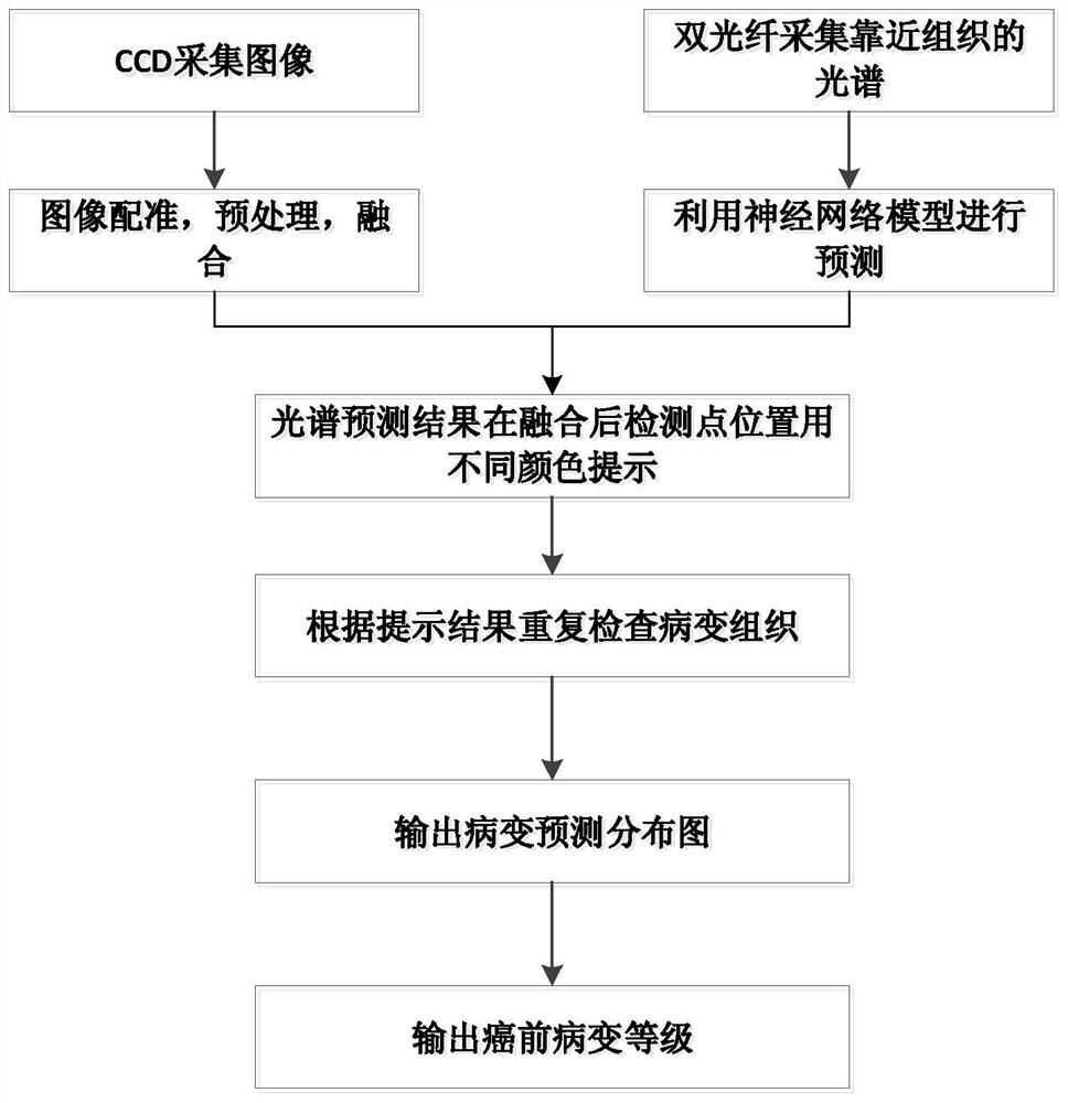
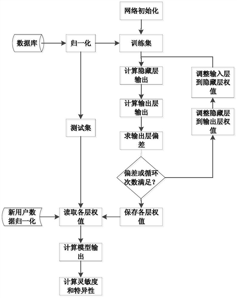
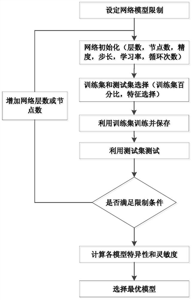
[0030] The invention relates to a method for screening precancerous lesions of cervical cancer, which uses spectral extraction features to establish a neural network model to classify precancerous lesions, and utilizes image fusion technology to locate transitional zones of the cervix. The spectral detection results are displayed on the transition zone image, and different levels are represented by different color levels, which provides a more intuitive display for medical workers and provides guidance for key inspections and biopsies of lesion areas.

[0031] The data used in this method are divided into five categories, whose labels are normal, CINⅠ, CINⅡ, CINⅢ and cervical erosion. The data come from hospital clinical trials, and the labels are taken from the results of histopathological examination.

[0032] figure 1 It is a schematic diagram of the overall flo...


PUM


Abstract

The invention relates to a screening method for cervical precancerous lesions combining spectrum and images, and belongs to the fields of spectrum analysis and medical image processing. The present invention builds a database based on the spectral and image data collected simultaneously by the joint probe of the spectral and endoscope, uses the spectral database to construct a neural network precancerous lesion classification model, uses image registration and fusion, and uses different spectral diagnostic results of corresponding detection points. The color is displayed on the corresponding detection point image, reconstructed into a distribution map of precancerous lesions, and the diagnosis result is output according to the distribution map. The present invention can be used alone as a screening algorithm for cervical cancer, and can also provide image guidance for further biopsy. The diagnosis result of this method is a distribution map of cervical precancerous lesions, which is objective and direct, and is not affected by the technique of the operator.

Description

technical field [0001] The invention relates to a screening method for cervical precancerous lesions combining spectrum and images, and belongs to the fields of spectrum analysis and medical image processing. Background technique [0002] Cervical cancer is a common gynecological malignancy, which has caused serious harm to women's health. The cure rate of early cervical cancer can reach more than 90%, while the cure rate of carcinoma in situ is 100%. Early screening of cervical cancer can prevent the continued development of cervical cancer and prevent the occurrence of cervical cancer. Therefore, early diagnosis of cervical cancer is very important. Clinically, early lesions of cervical cancer are divided into CINI grade, CINII grade and CINIII grade according to the degree of cervical epithelial cell lesions, and invasive cancer is defined as CINIII grade or later. The development of early lesions to invasive cancer is slow, and timely and universal early screening is ...

Claims


Application Information

Patent Timeline
Patent Type & Authority Patents(China)
IPC IPC(8): A61B5/00
CPCA61B5/7264
Inventor 钱志余刘文文晋晓飞李俊俊张欢
Owner NANJING UNIV OF AERONAUTICS & ASTRONAUTICS
Learn More