
Auxiliary evaluation method for renal interstitial fibrosis, computer equipment and computer readable storage medium
A technology for renal interstitial fibrosis and fibrosis, which is applied in computing, neural learning methods, image data processing, etc., can solve problems such as low accuracy, inability to obtain parameter values, and increased time cost, and improve detection efficiency. , the effect of good robustness and generalization ability, good accuracy
- Summary
- Abstract
- Description
- Claims
- Application Information
AI Technical Summary
Problems solved by technology
Method used
Image

Examples
Embodiment 1
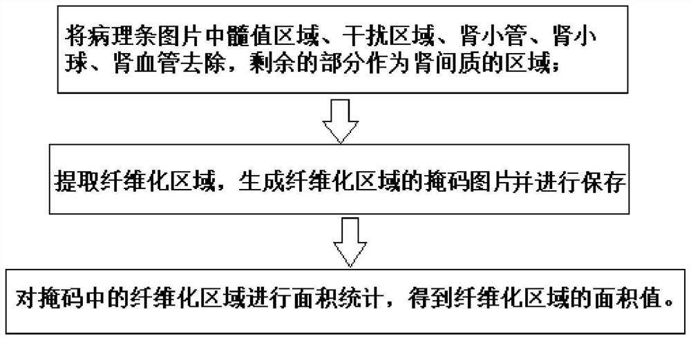
[0030] An auxiliary assessment method for renal interstitial fibrosis, comprising the following steps:
[0031] (1) Remove the medulla area, interference area, renal tubule, glomerulus, and renal blood vessels in the pathological strip picture, and the remaining part is used as the interstitial area of the kidney; the pathological strip picture is a picture scanned by a pathological slice scanner . In the present invention, the order of removing the medullary region, partially disturbed region, renal tubules, glomeruli, and renal blood vessels is not limited, as long as they are removed.
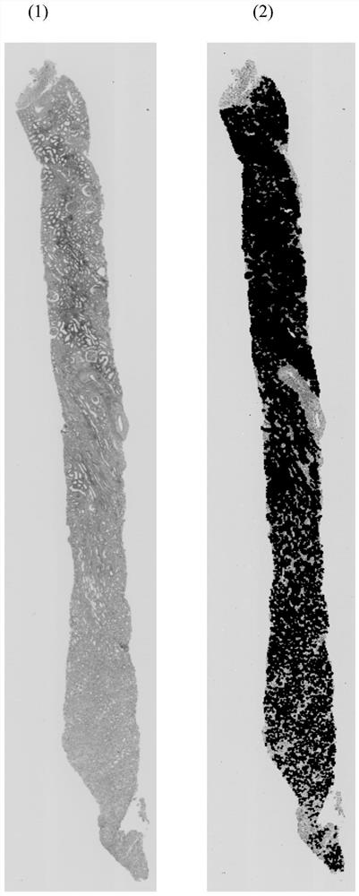
[0032] (2) Extract fibrosis area: After MASSON staining, the fibrosis area in the pathological strip presents a light green texture feature, which is extracted using the HSV color space in opencv, and the fibrosis area is extracted using the cv2.inRange function,
[0033] (3) The area value of the fibrosis region can be obtained by performing statistics on the area of the fibrosis regio...
Embodiment 2
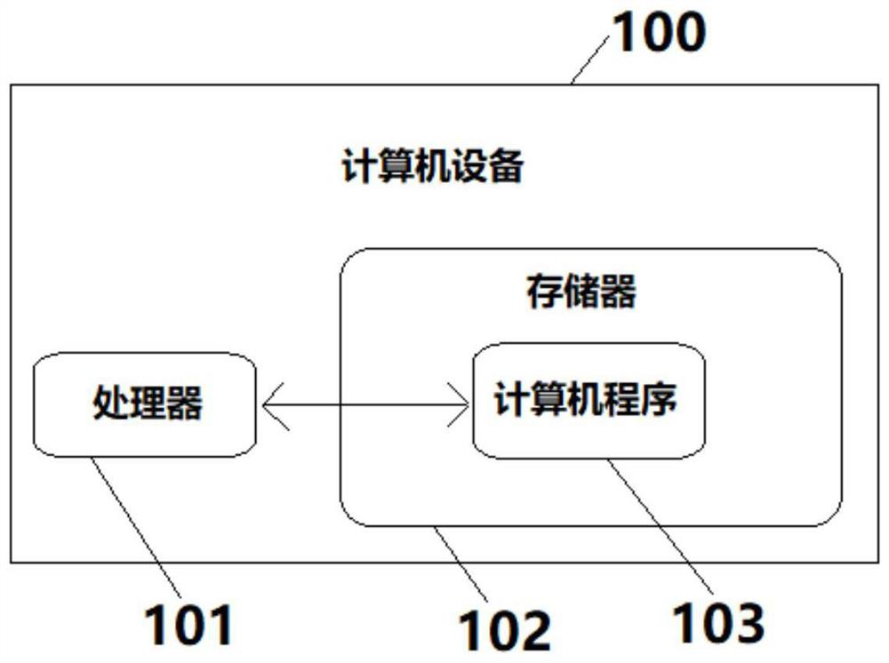
[0053] Embodiment 2, with reference to attached figure 2 .
[0054] In this embodiment, a computer device 100 is provided, including a memory 102, a processor 101, and a computer program 103 stored in the memory 102 and operable on the processor 101. When the processor 101 executes the computer program 103, it can realize The steps in the auxiliary assessment method for renal interstitial fibrosis provided in Example 1 above.
Embodiment 3
[0056] In this embodiment, a computer-readable storage medium is provided, on which a computer program is stored, and when the computer program is executed by a processor, the method for assisting assessment of renal interstitial fibrosis provided in the above-mentioned embodiments can be realized in the steps.
[0057] In this embodiment, the computer program may be the computer program in Embodiment 2.
[0058] In this embodiment, the computer-readable storage medium may be executed by the computer device in Embodiment 2.
PUM
 Abstract
Description
Claims
Application Information