Method of external constructing tissue engineering blood vessel
A tissue engineering and vascular technology, applied in the field of tissue engineering and materials science, can solve the problems of flat and inelastic lumen
- Summary
- Abstract
- Description
- Claims
- Application Information
AI Technical Summary
Problems solved by technology
Method used
Image
Examples
preparation example Construction
[0051] The preparation method of the tissue-engineered blood vessel of the present invention is simple. A certain number of smooth muscle cells are inoculated on a pharmaceutically acceptable biodegradable material, and then the cell-material compound is wrapped in an elastic material tube; Conditionally cultivate the cell material complex of step (b) for 3-10 days (more preferably 5-8 days); then apply the following dynamic stimulation through the elastic material tube, and continue to cultivate for 5-50 days (preferably 7-30 days) day): beat frequency: 75±10 beats / min; dilatation: 5±1%; pressure: 0.001-0.04Mpa; flow: 0.01-100ml / min, thereby obtaining tissue-engineered vascular grafts.
[0052] The shape of the tissue engineered vascular graft of the present invention is usually ring. The thickness of the vascular graft of the present invention is not particularly limited, and is usually 5-200 microns, preferably 10-50 microns, more preferably 20-40 microns.
[0053] The smo...
Embodiment 1
[0063] Isolation, culture, expansion and observation of vascular smooth muscle cells
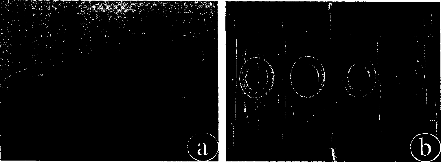
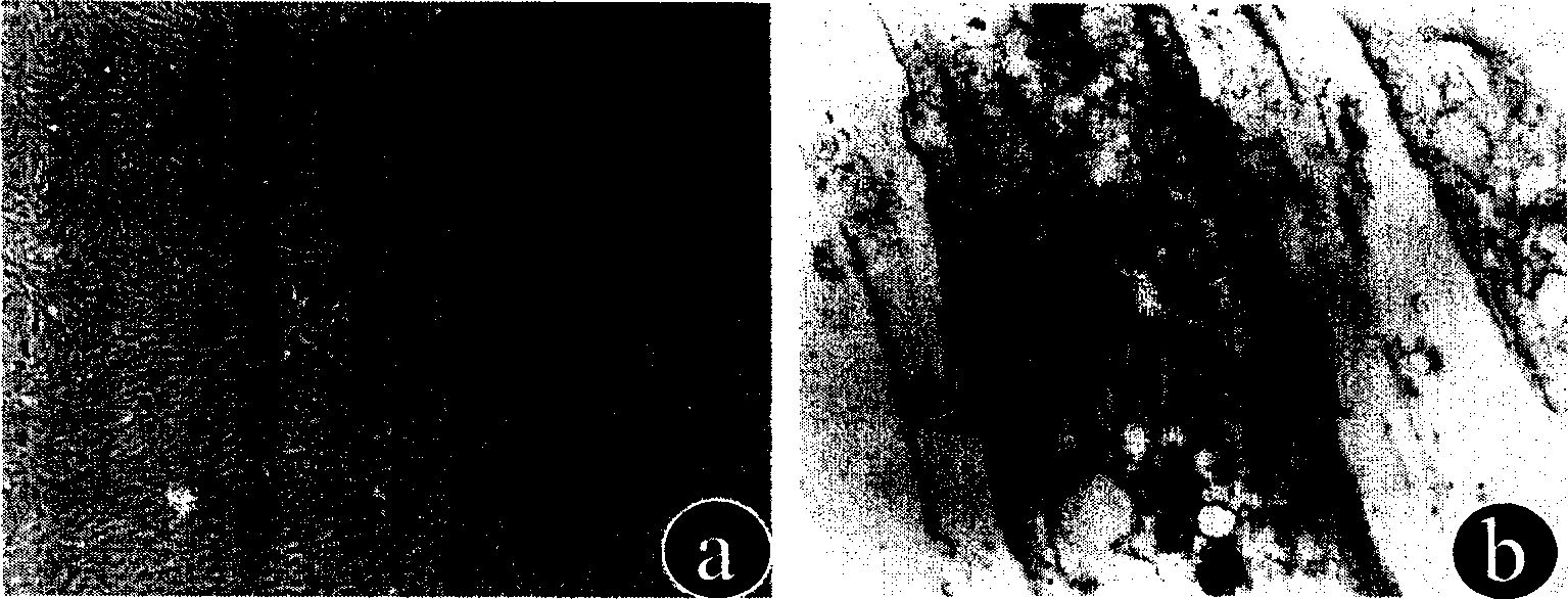
[0064] Select young Changfeng hybrid pigs, male or female, weighing 10-15kg, aged 2 months. Under a sterile environment, a section of porcine common carotid artery was surgically excised, 3-4cm long, after peeling off the adventitia and scraping off the intima, the arterial media was cut into 1×1×1mm 3 Spread the tissue pieces evenly on the culture dish, add DMEM culture solution containing 10% fetal bovine serum (FBS) after adhering to the wall, place at 37°C, 5% CO 2 The cells were cultured in an incubator, and the second generation cells were used for experiments. Cell growth was observed with an inverted phase-contrast microscope, and samples were taken for immunohistochemical detection of the expression of α-smooth muscle actin, and its ultrastructure was observed with a transmission electron microscope.
[0065] result:
[0066] (a) Morphological observation of cells
[0067] On th...
Embodiment 2
[0073] Smooth muscle cell identification
[0074] Immunohistochemical staining for detection of α-smooth muscle actin: smooth muscle cell slides were divided into experimental group and blank control group, and human skin fibroblast slides were retained as negative control group. Main steps Tissue sections were fixed in anhydrous acetone, treated with 0.25% TritonX-100 to increase cell permeability, treated with 1.5% H2O2 to block endogenous peroxidase, incubated with sheep serum to remove non-specific staining, and dropped primary antibody (mouse Anti-human smooth muscle α-actin antibody diluted 1:50 (DAKO company, Denmark), PBS was added dropwise to the blank control group, 4 ° C refrigerator overnight, and the secondary antibody (horseradish peroxidase-labeled goat anti-mouse IgG) was added dropwise. React at 37°C for 30 minutes, develop color with DAB, stain with hematoxylin, dehydrate with gradient alcohol, and seal with neutral resin. Judgment of the results: the cells ...
PUM
| Property | Measurement | Unit |
|---|---|---|
| thickness | aaaaa | aaaaa |
Abstract
Description
Claims
Application Information
 - R&D
- Intellectual Property
- Life Sciences
- Materials
- Tech Scout
- Unparalleled Data Quality
- Higher Quality Content
- 60% Fewer Hallucinations
Browse by: Latest US Patents, China's latest patents, Technical Efficacy Thesaurus, Application Domain, Technology Topic, Popular Technical Reports.
© 2025 PatSnap. All rights reserved.Legal|Privacy policy|Modern Slavery Act Transparency Statement|Sitemap|About US| Contact US: help@patsnap.com


