Realtime four-dimensional electro cardiogram imaging method and device

An electrocardiogram, four-dimensional technology, used in medical science, sensors, diagnostic recording/measurement, etc.

Inactive Publication Date: 2006-08-16
张士东
View PDF1 Cites 9 Cited by
  • Summary
  • Abstract
  • Description
  • Claims
  • Application Information

AI Technical Summary

Problems solved by technology

It uses computer imaging technology to describe the three-dimensional space structure of a single ECG vector, but because the three-dimensional space description method it uses is from the origin as the starting point to the display method of the three-dimensional ECG vector value as the end point, the starting point of the ECG vector must be It is a point with zero potential on the X, Y, and Z axes, so it is impossible to display various ECG vectors (including abnormal ECG vectors and a large number of normal ECG vectors) whose starting point is not zero potential in three-dimensional space, and real-time show

Method used


Image

  • Realtime four-dimensional electro cardiogram imaging method and device
  • Realtime four-dimensional electro cardiogram imaging method and device
  • Realtime four-dimensional electro cardiogram imaging method and device

Examples


Embodiment Construction

[0050] The following will be explained in conjunction with the accompanying drawings

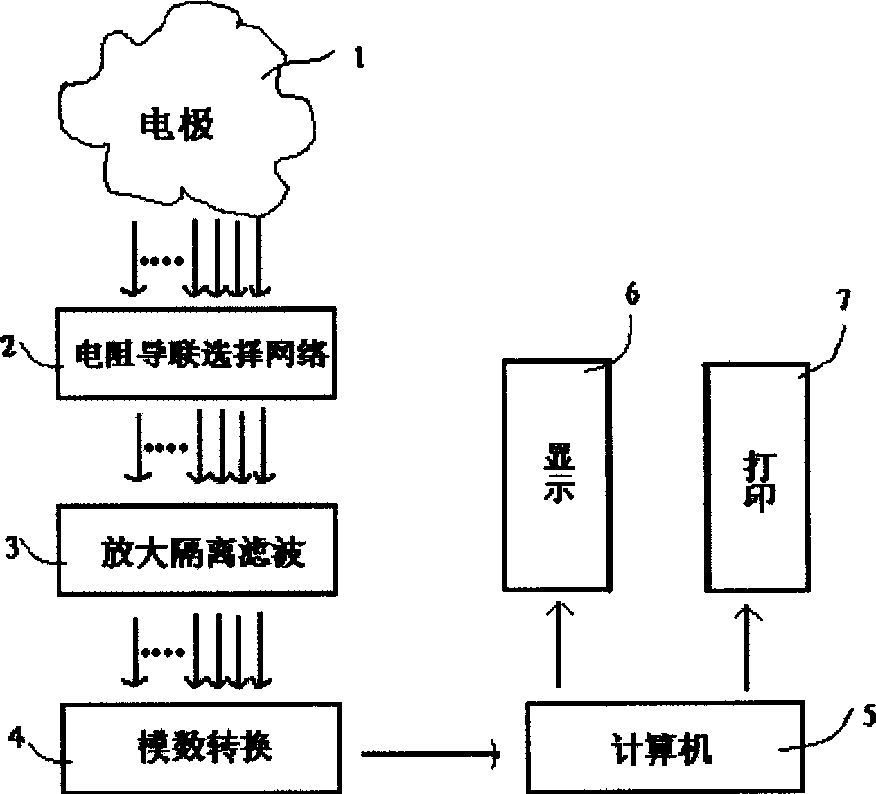
[0051] figure 1 The electrode 1 represents the Wilson electrode and the Frank electrode. The electrocardiographic signal of the human body is converted into a digital signal through the resistance lead selection network circuit 2, the amplification isolation filter circuit 3 and the analog-to-digital conversion circuit 4, and then input into the computer 5 . Using VC++ language, using computer window technology, real-time digital signal processing technology, computer three-dimensional imaging technology, database technology, the digitized human ECG signal is stored and processed by computer to form a four-dimensional electrocardiogram, 12-lead electrocardiogram, high-frequency electrocardiogram, Frequency-domain electrocardiogram, Q-T dispersion, heart rate variability, etc., are displayed by the monitor 6 or output by the printer 7.

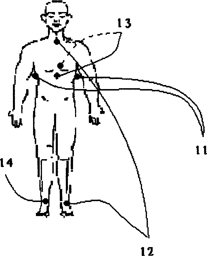
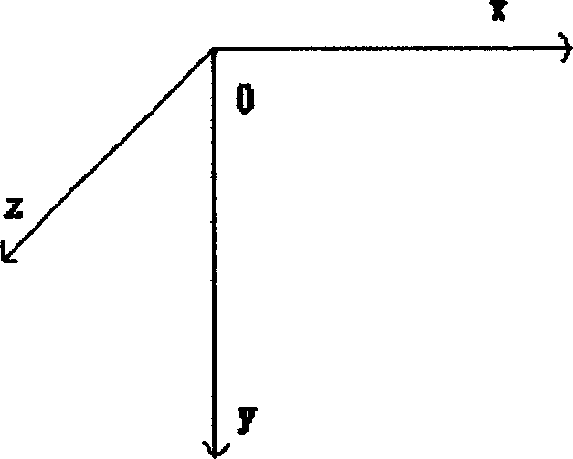
[0052] Fig. 2 shows how the three pairs of mutually pe...


PUM


Abstract

A real-time 4D electrocardiogram imaging method and its apparatus are disclosed. The mutually perpendicular three pairs of electrodes are arranged around heart to form a 3D space. Their intersection point is the origin point of said 3D space. Said three pairs of electrodes express X, Y and Z axises and time expressed the fourth dimension. The instantaneous cardioelectric voltages recorded by three pairs of electrodes form the instantaneous position of 3D cardioelectric vector. The continuous instantaneous positions are linked by lines to form the 4D electrocardiogram, which is dynamically displayed by computer.

Description

technical field [0001] The invention relates to a four-dimensional electrocardiogram imaging method and device. It belongs to medical electronic equipment. Background technique [0002] It is currently known that the electrical activity of the human heart is based on the following: a. The ion concentration difference inside and outside the cardiomyocyte forms the potential difference between the inside and outside of the cell membrane, the outside of the cell is positive charge, and the inside of the cell is negative charge. At this time, the potential difference between the inside and outside of the cell membrane is in a resting state, and no action potential is generated. called repolarization. b. When the ion concentration inside and outside the cell changes, it causes a change in the potential difference between the inside and outside of the cell membrane. The outside of the cell is negatively charged, and the inside of the cell is positively charged. This is called de...

Claims


Application Information

Patent Timeline
Patent Type & Authority Applications(China)
IPC IPC(8): A61B5/0402
Inventor 吴立群张士东
Owner 张士东
Why Eureka
Try Eureka