Injectable glaucoma implants with multiple openings

a glaucoma and implant technology, applied in the field of shunt-type stenting devices, can solve the problems of significant side effects, blindness if untreated, and patients may suffer substantial, irreversible vision loss, etc., and achieves rapid visual recovery, less morbidity, and faster surgical procedures.

Active Publication Date: 2005-12-08
GLAUKOS CORP
View PDF105 Cites 276 Cited by
  • Summary
  • Abstract
  • Description
  • Claims
  • Application Information

AI Technical Summary

Benefits of technology

[0026] Glaucoma surgical morbidity would greatly decrease if one were to bypass the focal resistance to outflow of aqueous only at the point of resistance, and to utilize remaining, healthy aqueous outflow mechanisms. This is in part because episcleral aqueous humor exerts a backpressure that prevents intraocular pressure from falling too low, and one could thereby avoid hypotony. Thus, such a surgery may virtually eliminate the risk of hypotony-related maculopathy and choroidal hemorrhage. Furthermore, visual recovery would be very rapid, and the risk of infection may be very small, reflecting a reduction in incidence from 2-5% to about 0.05%.
[0028] One technique performed in accordance with certain aspects herein can be referred to generally as “trabecular bypass surgery.” Advantages of this type of surgery include lowering intraocular pressure in a manner which is simple, effective, disease site-specific, and can potentially be performed on an outpatient basis.
[0036] Some embodiments provide a self-trephining glaucoma stent and methods thereof which advantageously allow for a “one-step” procedure in which the incision and placement of the stent are accomplished by a single device and operation. This desirably allows for a faster, safer, and less expensive surgical procedure. In any of the embodiments, fiducial markings, indicia, or the like and / or positioning of the stent device in a preloaded applicator may be used for proper orientation and alignment of the device during implantation.
[0037] Among the advantages of trabecular bypass surgery is its simplicity. The microsurgery may potentially be performed on an outpatient basis with rapid visual recovery and greatly decreased morbidity. There is a lower risk of infection and choroidal hemorrhage, and there is a faster recovery, than with previous techniques.
[0039] Some embodiments disclosed herein relate to a “foldable” stent wherein the size of the stent is reduced in order to place it through a yet smaller ocular entrance wound, as small as half or less than the size of the unfolded stent. The small wound size aids in recovery, to reduce the likelihood of complications, and to reduce the preparation and extent of the surgical environment. In another embodiment, the device is positioned through the trabecular meshwork in an ab externo or ab interno procedure. Reliable visualization (OCT, UBM, gonioscope, electromagnetic or other means) is a key enabler for micro precision surgery such as a trabecular bypass surgery using a microstent.

Problems solved by technology

Glaucoma causes pathological changes in the optic nerve, visible on the optic disk, and it causes corresponding visual field loss, resulting in blindness if untreated.
Patients may suffer substantial, irreversible vision loss prior to diagnosis and treatment.
However, there are secondary open-angle glaucomas which may include edema or swelling of the trabecular spaces (e.g., from corticosteroid use), abnormal pigment dispersion, or diseases such as hyperthyroidism that produce vascular congestion.
However, these drug therapies for glaucoma are sometimes associated with significant side effects, such as headache, blurred vision, allergic reactions, death from cardiopulmonary complications, and potential interactions with other drugs.
However, long-term review of surgical results showed only limited success in adults.
In retrospect, these procedures probably failed due to cellular repair and fibrosis mechanisms and a process of “filling in.” Filling in is a detrimental effect of collapsing and closing in of the opening created in the trabecular meshwork.
Once the openings close, the pressure builds back up and the surgery fails.
However, the relatively small hole created by this trabeculopuncture technique exhibits a filling-in effect and fails.
This was demonstrated not to succeed by clinical trial.
Although morbidity was zero in both trials, success rates did not warrant further human trials.
Failure was again from filling in of surgically created defects in the trabecular meshwork by repair mechanisms.
Neither of these is a viable surgical technique for the treatment of glaucoma.
The risk of placing a glaucoma drainage device also includes hemorrhage, infection, and diplopia (double vision).
All of the above surgeries and variations thereof have numerous disadvantages and moderate success rates.
They involve substantial trauma to the eye and require great surgical skill in creating a hole through the full thickness of the sclera into the subconjunctival space.
However, modifying existing filtering surgery techniques in any profound way to increase their effectiveness appears to have reached a dead end.

Method used


Image

  • Injectable glaucoma implants with multiple openings
  • Injectable glaucoma implants with multiple openings
  • Injectable glaucoma implants with multiple openings

Examples


Embodiment Construction

[0150] The preferred embodiments described herein relate particularly to surgical and therapeutic treatment of glaucoma through reduction of intraocular pressure and / or stimulation of the trabecular meshwork tissue. While the description sets forth various embodiment-specific details, it will be appreciated that the description is illustrative only and should not be construed in any way as limiting the inventions disclosed herein. Furthermore, various applications of the inventions disclosed herein, and modifications thereto, which may occur to those who are skilled in the art, are also encompassed by the general concepts described herein.

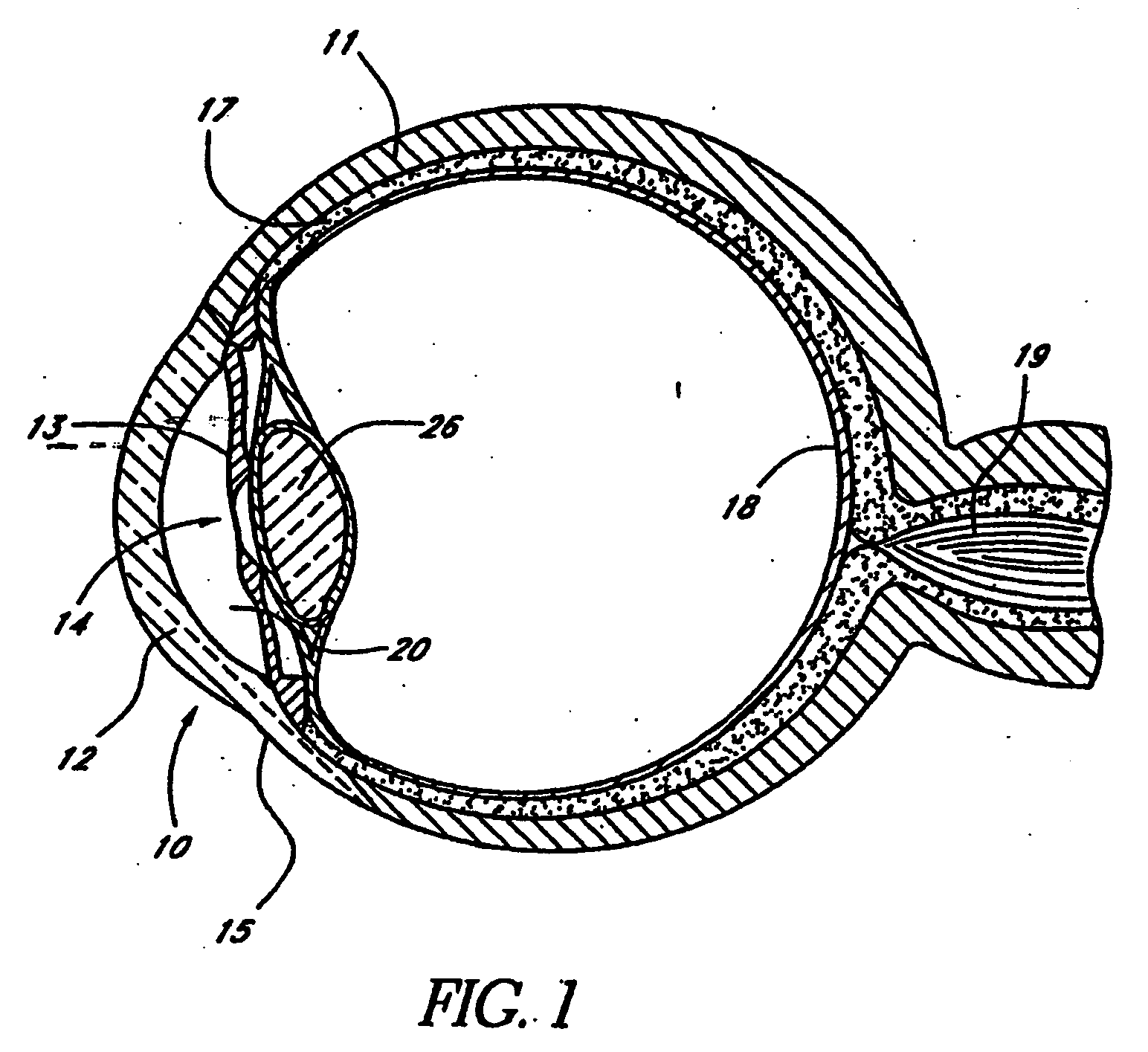
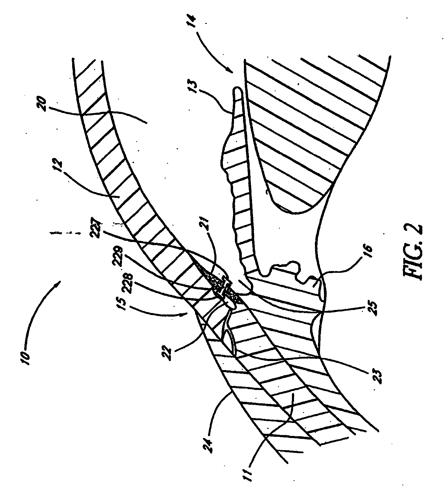
[0151]FIG. 1 is a cross-sectional view of an eye 10. FIG. 2 is an enlarged sectional view of the eye showing the relative anatomical locations of a trabecular meshwork 21, an anterior chamber 20, and a Schlemm's canal 22. A sclera 11 is a thick collagenous tissue which covers the entire eye 10 except a portion which is covered by a cornea 12.

[015...


PUM


Abstract

Intraocular stents and applicators are disclosed for treating glaucoma. The stents are configured to extend between the anterior chamber of the eye and Schlemm's canal for enhancing outflow of aqueous from the anterior chamber so as to reduce intraocular pressure. The stents can have features for anchoring the stent into Schlemm's canal as well as preventing the walls of Schlemm's canal from closing the outlet of the stents. The applicators can be steerable so as to make implantation easier. Additionally, the applicators can be configured to hold a plurality of stents so that multiple stents can be implanted through one incision without removing the applicator from the incision between serial implantations.

Description

BACKGROUND OF THE INVENTIONS [0001] 1. Field of the Inventions [0002] The present application relates generally to medical devices and methods for reducing the intraocular pressure in an animal eye and, more particularly, to shunt-type stenting devices for permitting and / or enhancing aqueous outflow from the eye's anterior chamber toward existing outflow pathways and associated methods thereof for the treatment of glaucoma in general. [0003] 2. Description of the Related Art [0004] The human eye is a specialized sensory organ capable of light reception and able to receive visual images. The trabecular meshwork serves as a drainage channel and is located in the anterior chamber angle formed between the iris and the cornea. The trabecular meshwork maintains a balanced pressure in the anterior chamber of the eye by allowing aqueous humor to flow from the anterior chamber. [0005] About two percent of people in the United States have glaucoma. Glaucoma is a group of eye diseases encompas...

Claims


Application Information

Patent Timeline
Patent Type & Authority Applications(United States)
IPC IPC(8): A61F2/00A61F2/14A61F9/007
CPCA61F9/00781A61M27/002A61M27/006
Inventor TU, HOSHENGARTOF, JASONHAFFNER, DAVID
Owner GLAUKOS CORP
Why Eureka
Try Eureka