Ocular fundus portion analyzer and ocular fundus portion analyzing method

a technology of ocular fundus and analyzer, which is applied in the field of ocular fundus portion analyzer, can solve the problems of difficult to detect excavation in an early stage and accurately and quickly diagnose diseased areas, and achieve accurate and quick detection and diagnosis, and the effect of precise and quick perception

Inactive Publication Date: 2007-08-16
YOKOHAMA TLO
View PDF3 Cites 12 Cited by
  • Summary
  • Abstract
  • Description
  • Claims
  • Application Information

AI Technical Summary

Benefits of technology

[0024] According to the present invention, a shape of an optic disk, retinal and other ocular fundus shape can be precisely and quickly perceived, so that accurate and quick detection and diagnoses of ocular fundus defects, such as an excavation of an optic disk due to glaucoma and a macula retinae due to retinal detachment, become possible even from a large number of single-shot ocular fundus images; thus, remarkable effects can be obtained in the welfare.

Problems solved by technology

Therefore, it has been difficult to detect an excavation in an early stage and diagnose diseased areas accurately and quickly.

Method used


Image

  • Ocular fundus portion analyzer and ocular fundus portion analyzing method
  • Ocular fundus portion analyzer and ocular fundus portion analyzing method
  • Ocular fundus portion analyzer and ocular fundus portion analyzing method

Examples


examples

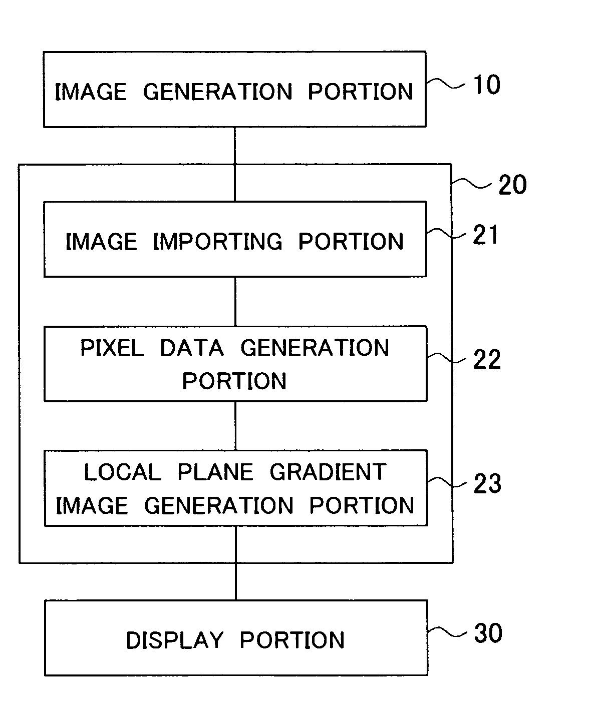
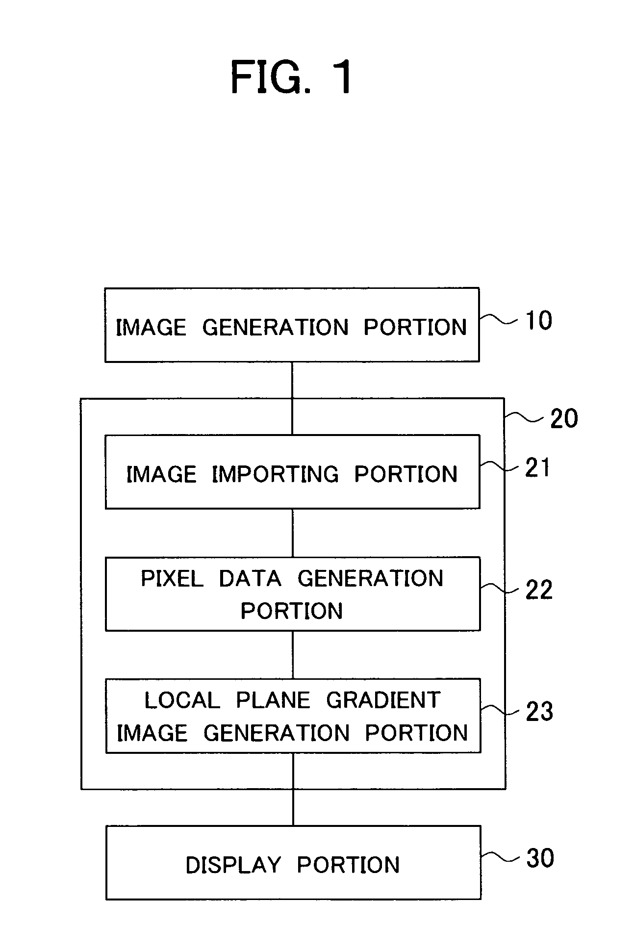
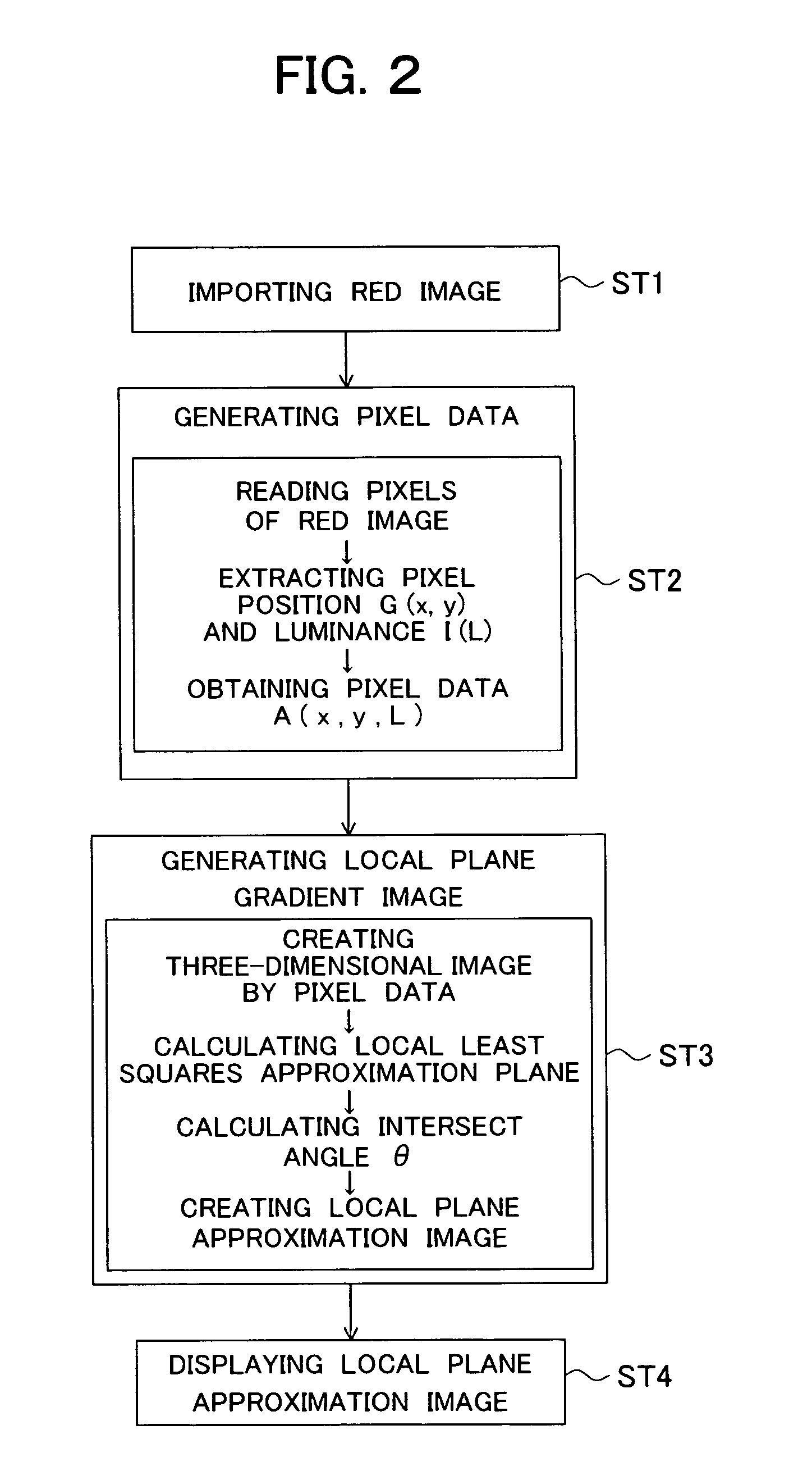
[0070] Below, the present invention will be explained in detail by using FIG. 4 to FIG. 9.

[0071]FIG. 4 is a red image of an optic disk of a healthy person, and FIG. 5 shows a red image in the case of an affected person. FIG. 6 is a three-dimensional image of an optic disk of the healthy person created from FIG. 4 by using the present invention, and FIG. 7 is that of the affected person and corresponds to FIG. 5. FIG. 8 expresses as a contour drawing a local plane gradient image of a healthy person based on a gradient angle corresponding to pixel positions, and FIG. 9 expresses that of the affected person.

[0072] The red image of an optic disk of a healthy person (FIG. 4) obtained by an ocular fundus examination and the red image of an optic disk of an affected person (FIG. 5) were imported to the image analyzer, pixel positions and luminance were measured to create pixel data, and the result was recorded in a memory device. The recorded pixel data was read to a stereoscopic image d...


PUM


Abstract

An ocular fundus portion analyzer and the method are provided for attaining accurate and quick detection and diagnoses of an ocular fundus defect portion, such as an excavation of an optic disk caused by glaucoma and a macula retinae caused by retinal detachment, by dealing with a large number of single-shot ocular fundus images, for example, taken in physical examinations. It is configured to include an image importing portion for importing an image of an ocular fundus portion taken by a visible radiation or a near-infrared radiation; a pixel data generation portion for generating pixel data including pixel positions and luminance of pixels constituting the image; a local plane gradient image generation portion for generating a three-dimensional image from pixel positions and luminance of the pixel data, obtaining a local approximation plane at any pixel and generating a local plane gradient image from a gradient angle with respect to a control plane of the three-dimensional image; and a display portion for displaying the local plane gradient image.

Description

TECHNICAL FIELD [0001] The present invention relates to an ocular fundus portion analyzer and an ocular fundus portion analyzing method, and particularly relates to an ocular fundus portion analyzer and an ocular fundus portion analyzing method for surely detecting an excavation of an optic disk as an area affected by glaucoma, a macula retinae, retrobulbar optic neuritis and maculopathy due to retinal detachment, etc. from an ocular fundus photograph taken in an examination of an ocular fundus for finding eye diseases, such as glaucoma and retinal detachment in particular. Background Art [0002] It is known that, when suffered by glaucoma, an excavation seen as a pale area arises at an optic disk of an ocular fundus; and an existence and stage of the disease are diagnosed by detecting the same. To detect the excavation, a method that a doctor directly observes an ocular fundus image to diagnose and a method that a doctor observes a taken ocular fundus photograph of an affected perso...

Claims


Application Information

Patent Timeline
Patent Type & Authority Applications(United States)
IPC IPC(8): A61B3/14A61B3/12
CPCA61B3/14A61B3/12
Inventor TAJIMA, FUMIAKIMIYATAKE, NAOKISATO, HIDEAKIMATSUDA, NOBUO
Owner YOKOHAMA TLO
Why Eureka
Try Eureka