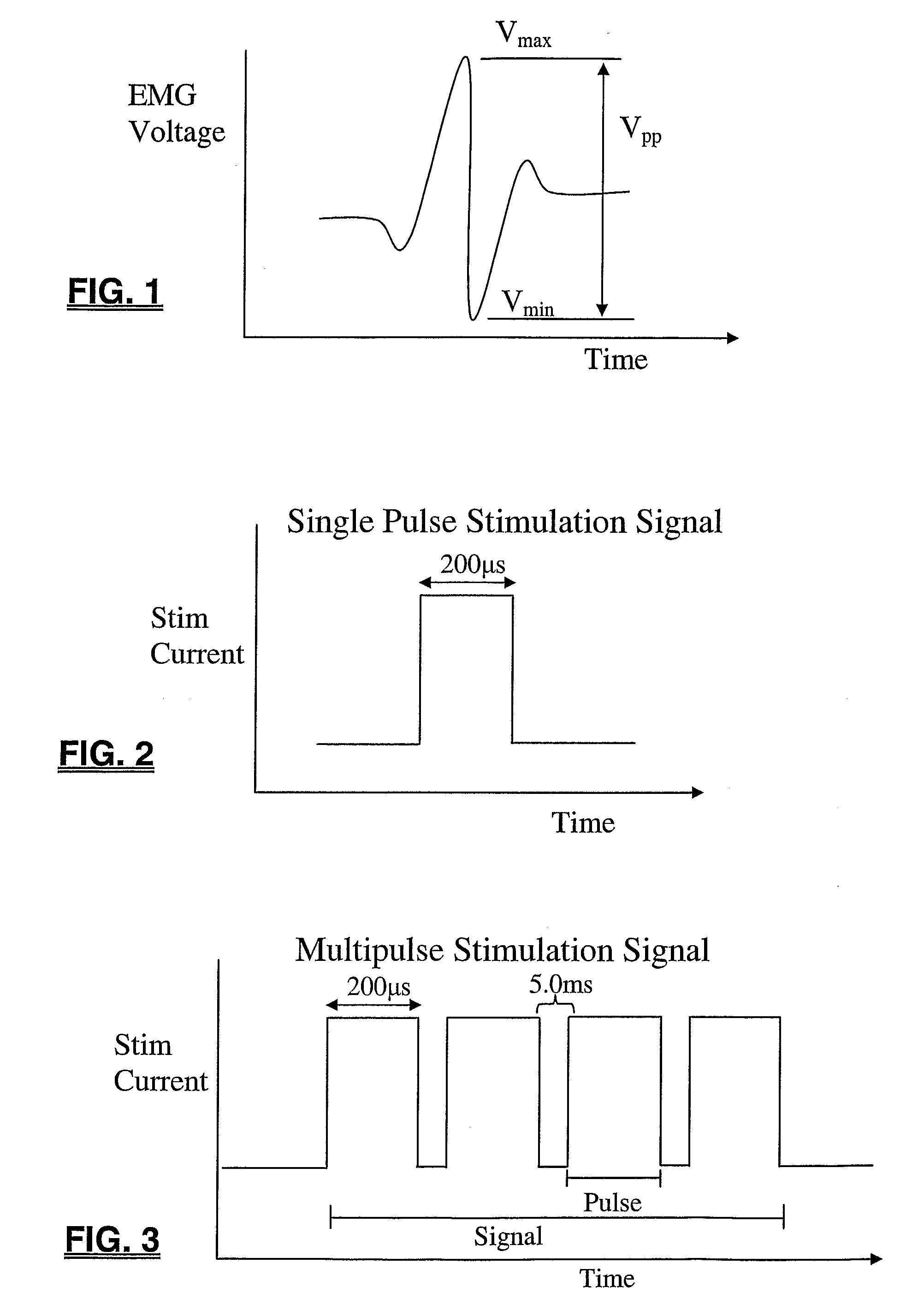
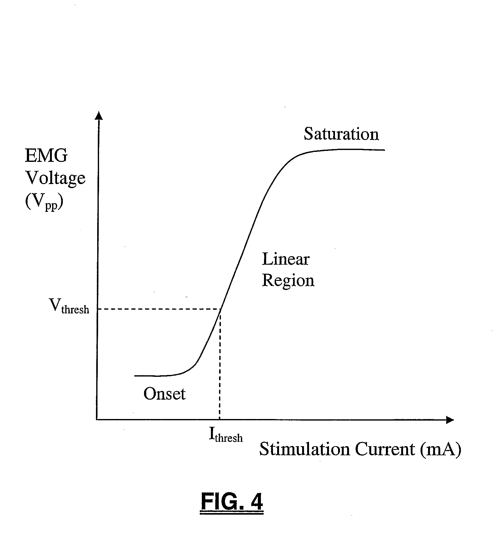
[0006]The present invention endows surgeons with valuable information that allows for the efficient assessment of risk to neural tissue before, during, and / or after a surgical procedure. This is accomplished by quickly and accurately determining a stimulation threshold for neural tissue and relaying that information to the surgeon in a simple comprehensible fashion. Stimulation thresholds are determined by electrically stimulating nerve tissue and analyzing resulting muscle activity relative to determine the stimulation current level at which nerve tissue depolarizes. To make stimulation threshold determinations, muscle activity may be monitored by measuring electrical signals associated with muscle contraction, called electromyography (“EMG”). EMG responses can be characterized by a peak-to-peak voltage of Vpp=Vmax−Vmin. Characteristics of the electrical stimulation signal used may vary depending upon several factors, including the particular nerve assessment performed, the spinal target level, the type of neural tissue stimulated (e.g. nerve root, spinal cord, brain, etc. . . ) among others.
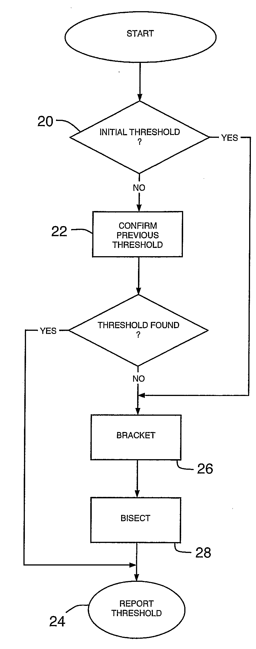
[0008]The algorithm described herein may considerably reduce the number of stimulations, and thus time, required to determine Ithresh, particularly for a number of channels, over the course of a procedure. The basic method for finding Ithresh utilizes a bracketing method and a bisection method. The bracketing method quickly finds a range (bracket) of stimulation currents that must contain Ithresh and the bisection method narrows the bracket until Ithresh is known within a specified accuracy.
[0011]To reduce the number of stimulations required to complete the bracketing and bisection steps when Ithresh is determined repeatedly and / or over multiple channels, the algorithm omits stimulations for which the result is predictable from data acquired during previous stimulations. When a stimulation is omitted, the algorithm proceeds as if the stimulation had taken place. However, instead of reporting an actual recruitment result, the reported result is inferred from the previous data. This permits the algorithm to proceed to the next step immediately, without the delay associated with a stimulation. For every stimulation signal delivered, the EMG response, or lack thereof, is detected and recorded on each channel (no matter which channel is actually being processed for Ithresh). Later the data can be referred back to, allowing the algorithm to omit a stimulation and infer whether or not the channel would recruit at the given stimulation current.
[0015]To further reduce the number of stimulations required to repeatedly find Ithresh over the course of a procedure, the algorithm includes a confirmation step. If Ithresh has been previously determined for a specific channel, the algorithm may simply confirm that Ithresh has not changed rather than beginning anew with the bracketing and bisection methods. The algorithm first determines whether it is conducting the initial threshold determination for the channel or whether there is a previous Ithresh determination. If it is not the initial determination, the algorithm confirms the previous determination. If the previous threshold is confirmed, the algorithm reports that value as the present Ithresh. If it is the initial Ithresh determination or if the previous threshold cannot be confirmed, the algorithm enters the bracketing and bisection states to determine Ithresh and then reports the value.
[0018]The algorithm described herein may be particularly useful when employed to monitor nerve pathology in conjunction with the use of a nerve retractor. A typical nerve retractor serves to pull or otherwise maintain a nerve outside the surgical corridor, thereby protecting the nerve from inadvertent damage or contact by the “active” instrumentation used to perform the actual surgery. While generally advantageous, it has been observed that such retraction can cause nerve function to become impaired or otherwise pathologic over time due to the retraction. Monitoring Ithresh during nerve retraction may be useful to assess the degree to which retraction of a nerve or neural structure affects the nerve function over time. One advantage of such monitoring is that the conduction of the nerve may be monitored during the procedure to determine whether the neurophysiology and / or function of the nerve changes (for the better or worse) as a result of the particular surgical procedure. For example, it may be observed that the nerve conduction decreases (indicated by an increase in Ithresh over time) during the retraction, indicating that the nerve function has been negatively affected. In contrast, the nerve conduction may increase (indicated by a decrease in Ithresh over time), indicating that the nerve function may have been restored or improved by the surgical procedure (such as during a successful decompression surgery, etc . . . ). As mentioned, a change in Ithresh may occur on any channel; therefore it is advantageous to calculate the actual Ithresh for each channel, as opposed to determining a value for just the channel with the highest or lowest Ithresh. The algorithm of the present invention accomplishes this while substantially limiting the number of stimulations required to do so. This may substantially reduce the time required to make an Ithresh determination which in turn may reduce the overall surgical time and risk to the patient.
[0019]The algorithm of the present invention may also be of particular use during Motor Evoked Potential (MEP) monitoring. When surgical procedures are performed in the proximity of the spinal cord, potential damage to the spinal cord is a paramount concern. Consequences of spinal cord damage may range from a slight loss of sensation to complete paralysis of the extremities, depending on the location and extent of damage. MEP monitoring, which generally involves monitoring the transmission of an electrical signal along the spinal cord, may be employed to assess the spinal cord before, during, and / or after surgery. Degradation or decreased conduction of an electrical signal, indicated by an increase in Ithresh, may indicate that the health of the spinal cord is compromised. Obtaining such information quickly may allow the surgeon to initiate corrective measures before the damage gets worse and / or becomes permanent. Similar to the nerve pathology monitoring mentioned above, changes in Ithresh indicating potential damage to the spinal cord may occur on any monitored channel, thus it is advantageous to calculate the actual Ithresh for each channel, as opposed to determining just the channel with the highest or lowest Ithresh. Employing the algorithm of the present invention again allows this to be done accurately and efficiently.

