Eureka AIR delivers breakthrough ideas for toughest innovation challenges, trusted by R&D personnel around the world.

Endoscopic tissue apposition device with multiple suction ports

a tissue apposition and suction port technology, applied in the field of endoscopic tissue apposition devices, can solve the problems of inability to properly function the valve at the junction between the stomach and the esophagus, long and cumbersome use of devices, and difficult surgical knots to tie successfully, etc., to facilitate the use of abrasion means, promote tissue ingrowth, and correct positioning of abrasions

Inactive Publication Date: 2008-09-04
CR BARD INC
View PDF99 Cites 3 Cited by
  • Summary
  • Abstract
  • Description
  • Claims
  • Application Information

AI Technical Summary

Benefits of technology

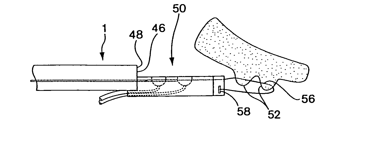
[0017]In the side-by-side tissue apposition embodiments, novel needle arrangements may be employed to penetrate tissue portions that are captured in the suction ports that are arranged away from the longitudinal axis of the capsule. A forked needle capable of simultaneously penetrating tissue portions held side-by-side is provided. For independent penetration of the tissue portions held captured in a side-by-side arrangement, a capsule design having a diverter in the needle track serves to guide independent needles to the selected suction port as they are advanced distally to penetrate tissue.
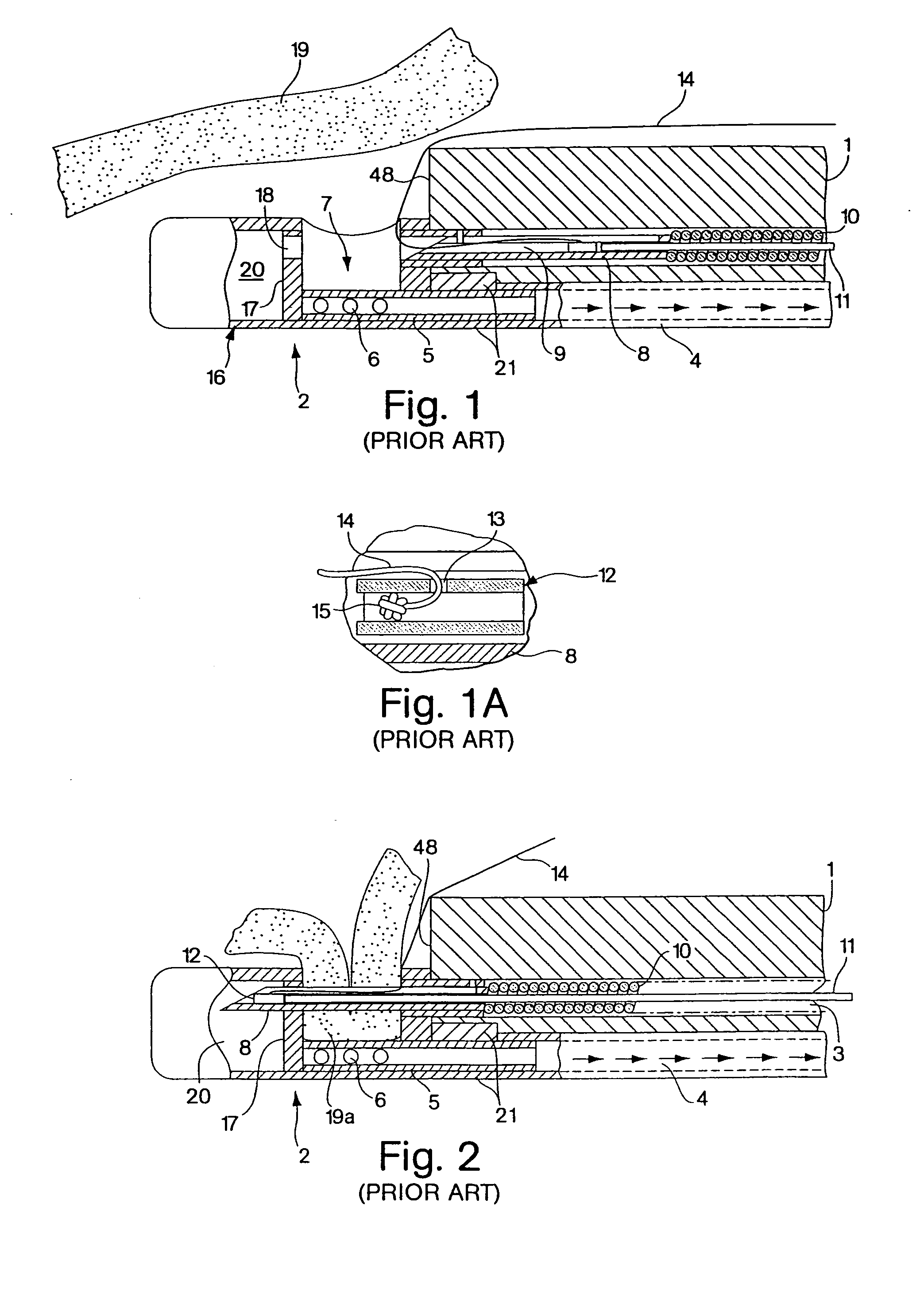
[0041]It is another object of the invention to provide an endoscopic tissue apposition device that safely abrades tissue sufficiently to initiate an injury response in that tissue without adversely and permanently damaging the tissue.

Problems solved by technology

This condition results from the inability of the valve at the junction between the stomach and the esophagus to function properly.
The use of the devices is, thus, long and cumbersome.
Surgical knots are difficult to tie successfully, especially for non-surgical physicians that may be performing the endoscopic suturing procedure.

Method used


Image

  • Endoscopic tissue apposition device with multiple suction ports
  • Endoscopic tissue apposition device with multiple suction ports
  • Endoscopic tissue apposition device with multiple suction ports

Examples


embodiment 200

[0121]With the side-by-side capsule embodiment 200, there are several possible mechanisms for tissue securement may be employed. Tissue securement may comprise suture material passed through the tissue portions. Alternatively, the tissue securement mechanism may comprise a clip that is driven into the tissue to secure the portions and remains in the patient, such as a helical wire coil described below. Several tissue securement advancement mechanisms are also possible. Specifically, several needle configurations and suture tag securement embodiments are possible with the multiple suction port capsule 200.

[0122]In one embodiment of the side-by-side device, shown in FIGS. 15-18, a forked needle 212 slidably mounted in the capsule 200 is employed to penetrate simultaneously the tissue portions captured in suction ports 202 for suture delivery. As best shown in the sectional isometric view of the capsule 200, shown in FIG. 18, the forked needle 212 may comprise a forked stainless steel ...

embodiment 400

[0134]FIG. 22 shows an alternate embodiment of a tissue apposition device having multiple suction ports that are longitudinally and angularly offset from each other. The offset capsule embodiment 400 may have a cylindrical shape having at least two suction ports 402 formed through the surface of the capsule that are spaced longitudinally on the capsule and angularly offset from each other. The exact placement of the ports on the suction chamber may be varied to obtain the preferred tissue plication shape for the given procedure. In the example of the offset capsule shown in FIG. 22, the suction ports 402 are arranged to be slightly spaced longitudinally of a distance of less than half the length of a suction port and are angularly displaced at less than 90°. This arrangement of suction ports offers a physician an alternative configuration of formed tissue plications that may better achieve the objective of the particular treatment. The arrangement of suction ports 402 is believed to...

embodiment 500

[0137]FIG. 24 shows another alternate embodiment of the multiple suction port tissue apposition device having four suction ports. The quad port apposition device 500 is similar to the side-by-side apposition device 300 described above, but comprises an additional set of two suction ports and two needles on the opposite side of the capsule. Additionally, the quad port embodiment 500 may have a cylindrical shape having a circular cross-section with suction ports 502 spaced around the circumference of the capsule. Preferably, each suction port 502 opens to an independent vacuum chamber 504 serviced by an independently operable needle 506. Preferably, the quad port capsule is used in conjunction with an endoscope 1 having multiple working channels 3 to accommodate the movement of the multiple needles 506. For example, an endoscope having two working channels 3 could accommodate two pusher cables 10 slidable within each working channel 3. Each pusher cable could then be joined to a separ...


PUM


Abstract

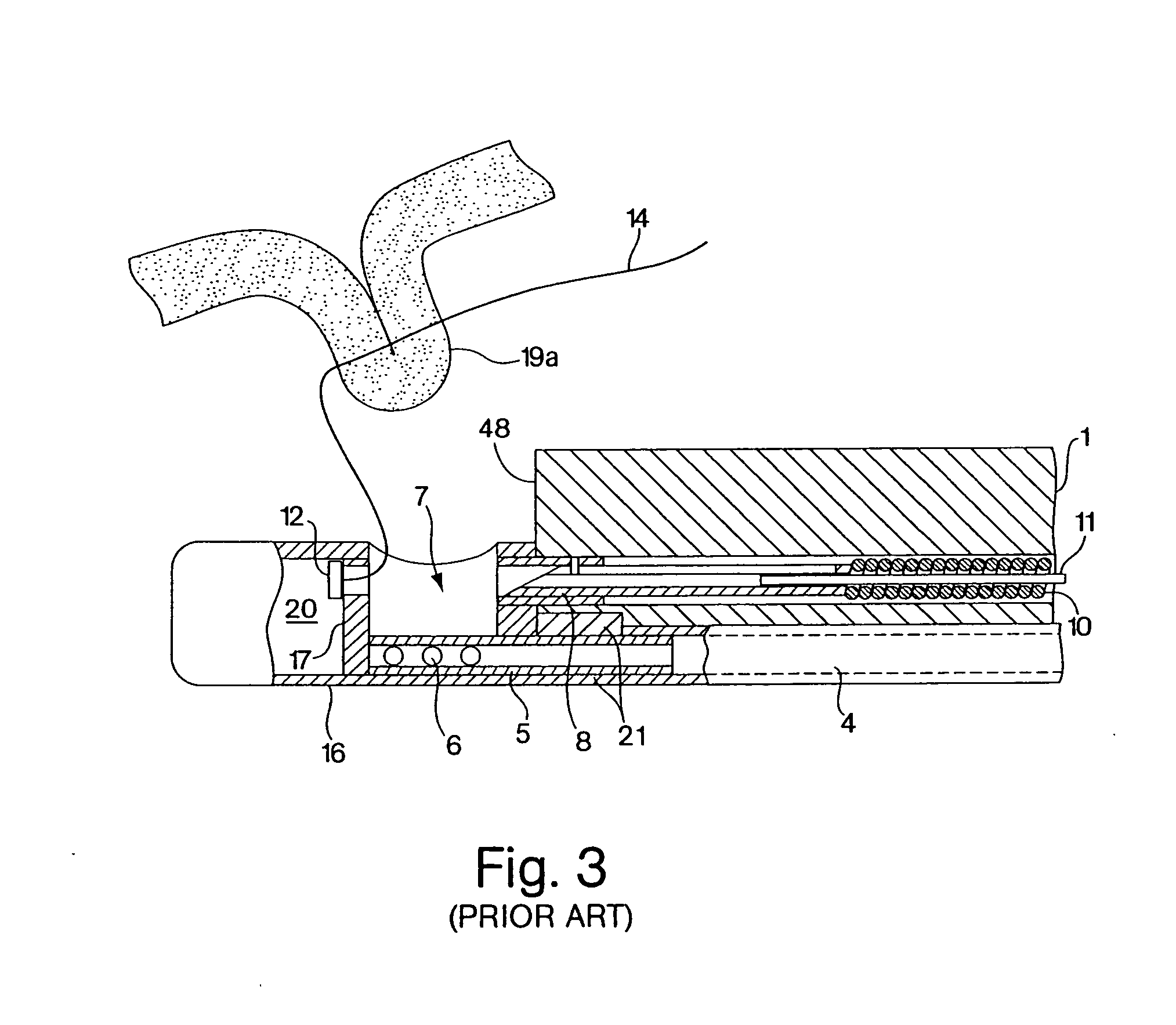
The present invention relates to an improved endoscopic tissue apposition device having multiple suction ports. The invention permits multiple folds of tissue to be captured in the suction ports with a single positioning of the device and attached together by a tissue securement mechanism such as a suture, staple or other form of tissue bonding. The improvement reduces the number of intubations required during an endoscopic procedure to suture tissue or join areas of tissue together. The suction ports may be arranged in a variety of configurations on the apposition device to best suit the desired resulting tissue orientation. The inventive tissue apposition device may also incorporate tissue abrasion means to activate the healing process on surfaces of tissue areas that are to be joined by operation of the device to promote a more secure attachment by permanent tissue bonding.

Description

RELATED APPLICATIONS[0001]This application is a continuation of U.S. application Ser. No. 10 / 220,379, filed Mar. 24, 2003, which is the U.S. National Stage of International Application No. PCT / US01 / 06835, filed Mar. 2, 2001, which claims the benefit to U.S. Provisional Applications Nos. 60 / 186,771, filed Mar. 3, 2000, 60 / 186,650, filed Mar. 3, 2000, and 60 / 187,275, filed Mar. 6, 2000, the entire contents of which are incorporated herein by reference.FIELD OF THE INVENTION[0002]The present invention relates to improvements for endoscopic tissue apposition devices. Specifically, the invention provides an endoscopic apposition device configured to collect a plurality of tissue portions with a single operation of the device so that the tissue can be joined together by a tissue securing means.BACKGROUND OF THE INVENTION[0003]Endoscopic apposition devices are devices that can be used in the body of a patient without the need to make an external incision in the patient, the device being co...

Claims


Application Information

Patent Timeline
Patent Type & Authority Applications(United States)
IPC IPC(8): A61B17/04A61B1/00A61B18/20A61B17/00A61B17/06A61B17/068A61B17/08A61B17/10A61B17/11A61B17/12A61B17/32A61B18/00A61B18/04A61B18/18A61M37/00
CPCA61B17/00234A61B2018/00291A61B17/0469A61B17/0482A61B17/0625A61B17/068A61B17/08A61B18/1492A61B18/22A61B2017/00827A61B2017/0409A61B2017/0417A61B2017/0458A61B2017/047A61B2017/0472A61B2017/061A61B2017/081A61B2017/306A61B17/0401
Inventor GAMBALE, RICHARD A.WEISER, MICHAEL F.PAGE, EDWARD C.LUKIN, PETER J.
Owner CR BARD INC
Why Eureka
Eureka Blog
Learn More