Endoscopic tissue apposition device with multiple suction ports

a tissue apposition and suction port technology, applied in the field of endoscopic tissue apposition devices, can solve the problems of inability to properly function the valve at the junction between the stomach and the esophagus, long and cumbersome use of devices, and difficult surgical knots to tie successfully, etc., to facilitate the use of abrasion means, promote tissue ingrowth, and correct positioning of abrasions

Inactive Publication Date: 2008-07-15
CR BARD INC
View PDF310 Cites 318 Cited by
  • Summary
  • Abstract
  • Description
  • Claims
  • Application Information

AI Technical Summary

Benefits of technology

[0027]The abrasion means may be incorporated in devices having a single suction port as those disclosed in the patents referenced above, but preferably the abrasion means is incorporated in an apposition device having multiple suction ports such as described herein. The arrangement and operation of the suction ports facilitates use of the abrasion means in that tissue can be held in place, in contact with the abrasion means, by applying light vacuum pressure through the vacuum chambers to collect tissue into the suction ports. By positioning the abrasion means adjacent the suction ports, tissue is reliably brought into contact with the abrasion means. Also facilitated is the correct positioning of the abrasion in the tissue area that will be brought into contact during the apposition procedure.
[0028]It is believed that joining area of tissue that have been injured slightly or abraded will undergo a healing process that promotes tissue ingrowth between the joined tissue surfaces resulting in a new, unified tissue mass rather than two separate tissues attached together by a tissue securement mechanism that could be subject to failure over time. Another advantage of the tissu

Problems solved by technology

This condition results from the inability of the valve at the junction between the stomach and the esophagus to function properly.
The use of the devices is, thus, l

Method used


Image

  • Endoscopic tissue apposition device with multiple suction ports
  • Endoscopic tissue apposition device with multiple suction ports
  • Endoscopic tissue apposition device with multiple suction ports

Examples


embodiment 200

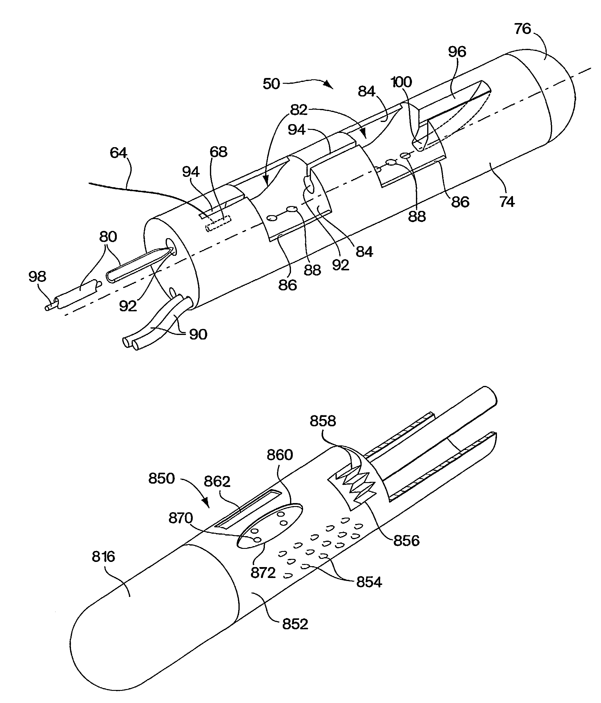
[0121]With the side-by-side capsule embodiment 200, there are several possible mechanisms for tissue securement may be employed. Tissue securement may comprise suture material passed through the tissue portions. Alternatively, the tissue securement mechanism may comprise a clip that is driven into the tissue to secure the portions and remains in the patient, such as a helical wire coil described below. Several tissue securement advancement mechanisms are also possible. Specifically, several needle configurations and suture tag securement embodiments are possible with the multiple suction port capsule 200.

[0122]In one embodiment of the side-by-side device, shown in FIGS. 15-18, a forked needle 212 slidably mounted in the capsule 200 is employed to penetrate simultaneously the tissue portions captured in suction ports 202 for suture delivery. As best shown in the sectional isometric view of the capsule 200, shown in FIG. 18, the forked needle 212 may comprise a forked stainless steel ...

embodiment 400

[0134]FIG. 22 shows an alternate embodiment of a tissue apposition device having multiple suction ports that are longitudinally and angularly offset from each other. The offset capsule embodiment 400 may have a cylindrical shape having at least two suction ports 402 formed through the surface of the capsule that are spaced longitudinally on the capsule and angularly offset from each other. The exact placement of the ports on the suction chamber may be varied to obtain the preferred tissue plication shape for the given procedure. In the example of the offset capsule shown in FIG. 22, the suction ports 402 are arranged to be slightly spaced longitudinally of a distance of less than half the length of a suction port and are angularly displaced at less than 90°. This arrangement of suction ports offers a physician an alternative configuration of formed tissue plications that may better achieve the objective of the particular treatment. The arrangement of suction ports 402 is believed to...

embodiment 500

[0137]FIG. 24 shows another alternate embodiment of the multiple suction port tissue apposition device having four suction ports. The quad port apposition device 500 is similar to the side-by-side apposition device 300 described above, but comprises an additional set of two suction ports and two needles on the opposite side of the capsule. Additionally, the quad port embodiment 500 may have a cylindrical shape having a circular cross-section with suction ports 502 spaced around the circumference of the capsule. Preferably, each suction port 502 opens to an independent vacuum chamber 504 serviced by an independently operable needle 506. Preferably, the quad port capsule is used in conjunction with an endoscope 1 having multiple working channels 3 to accommodate the movement of the multiple needles 506. For example, an endoscope having two working channels 3 could accommodate two pusher cables 10 slidable within each working channel 3. Each pusher cable could then be joined to a separ...


PUM


Abstract

An improved endoscopic tissue apposition device (50) having multiple suction ports (86) permits multiple folds of tissue to be captured in the suction ports (86) with a single positioning of the device (50) and attached together by a tissue securement mechanism such as a suture (14), staple or other form of tissue bonding. The improvement reduces the number of intubations required during an endoscopic procedure to suture tissue or join areas of tissue together. The suction ports (86) may be arranged in a variety of configurations on the apposition device (50) to best suit the desired resulting tissue orientation. The tissue apposition device (50) may also incorporate tissue abrasion means (852) to activate the healing process on surfaces of tissue areas that are to be joined by operation of the device to promote a more secure attachment by permanent tissue bonding.

Description

[0001]This application is the U.S. National stage of International Application No. PCT / US01 / 06835, filed on Mar. 2, 2001, published in English, claims the benefit of U.S. Provisional Application No. 60 / 186,771, filed on Mar. 2, 2000, U.S. Provisional Application No. 60 / 186,650, filed Mar. 3, 2000, and U.S. Provisional Application No. 60 / 187,275, filed Mar. 6, 2000. The entire teachings of the above applications are incorporated herein by reference.FIELD OF THE INVENTION[0002]The present invention relates to improvements for endoscopic tissue apposition devices. Specifically, the invention provides an endoscopic apposition devices configured to collect a plurality of tissue portions with a single operation of the device so that the tissue can be joined together by a tissue securing means.BACKGROUND OF THE INVENTION[0003]Endoscopic apposition devices are devices that can be used in the body of a patient without the need to make an external incision in the patient, the device being con...

Claims


Application Information

Patent Timeline
IPC IPC(8): A61B17/10A61B17/04A61B18/18A61B1/00A61B17/00A61B18/20A61B17/06A61B17/068A61B17/08A61B17/11A61B17/12A61B17/32A61B18/00A61B18/04A61M37/00
CPCA61B17/00234A61B17/0469A61B17/0625A61B17/068A61B17/08A61B17/0401A61B2018/00291A61B17/0482A61B18/1492A61B18/22A61B2017/00827A61B2017/0409A61B2017/0417A61B2017/0458A61B2017/047A61B2017/0472A61B2017/061A61B2017/081A61B2017/306
Inventor GAMBALE, RICHARD A.WEISER, MICHAEL F.PAGE, EDWARD C.LUKIN, PETER J.
Owner CR BARD INC
Why Eureka
Try Eureka