Imaging methods and apparatus particularly useful for two and three-dimensional angiography

a two-dimensional angiography and angiography technology, applied in angiography, medical science, diagnostics, etc., can solve the problem of inaccurate reference using a respiration sensor, and achieve the effect of enhancing the cnr of a dsa image and enabling diagnosis or treatmen

Inactive Publication Date: 2004-06-22
MEDIMAG C V I
View PDF22 Cites 17 Cited by
  • Summary
  • Abstract
  • Description
  • Claims
  • Application Information

AI Technical Summary

Benefits of technology

An object of the present invention is to provide an improved method and apparatus for producing two-dimensional or three-dimensional angiographic images to better enable diagnosis or treatment of a disease in the human body. A further object of the invention is to provide a method and apparatus for enhancing the CNR of a DSA image.
According to another aspect of the present invention, there is provided apparatus producing angiographical images of a portion of a patient's vascular system, comprising: a radiation source to be located at one side of the patient for radiating the portion of the patient's vascular system; a radiation detector to be located at the opposite side of the patient for producing electrical outputs corresponding to the magnitude of the radiation received by the detector; a computer for controlling the radiation source and for processing the outputs of the radiation detector to produce an image of the portion of the patient's vascular system; and a display for displaying the produced image; the computer controlling the radiation source and processing the outputs of the radiation detector such that: before a contrast material is injected into the patient's vascular system and a sequence of masking images is produced of the portion of the patient's vascular system, a determination is made as to the masking image in the sequence having a minimum change over its immediately preceding masking image; and after a contrast material is injected into the patient's vascular system and a corresponding sequence of contrast images is produced, from each contrast image the corresponding masking image in the sequence is subtracted starting with the masking image determined to have a minimum change over its immediately preceding masking image, to thereby produce a difference image having an enhanced contrast-to-noise ratio (CNR) which is displayed in the display.

Problems solved by technology

However, using a respiration sensor as the reference is not precise because of the difficulty in sharply defining a particular point of the respiration curve for this purpose.

Method used


Image

  • Imaging methods and apparatus particularly useful for two and three-dimensional angiography
  • Imaging methods and apparatus particularly useful for two and three-dimensional angiography
  • Imaging methods and apparatus particularly useful for two and three-dimensional angiography

Examples


Embodiment Construction

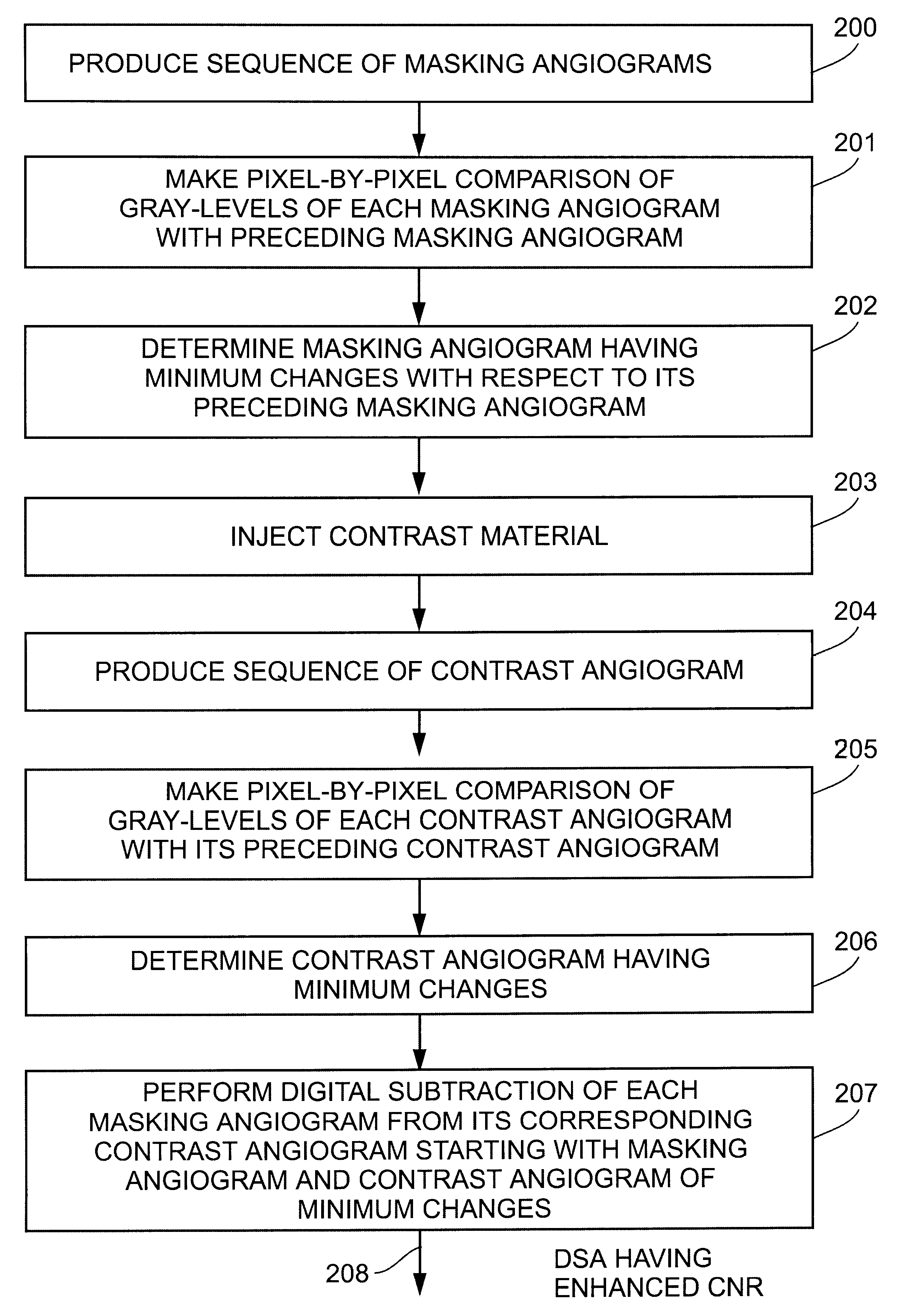
The Embodiment of FIGS. 1-6

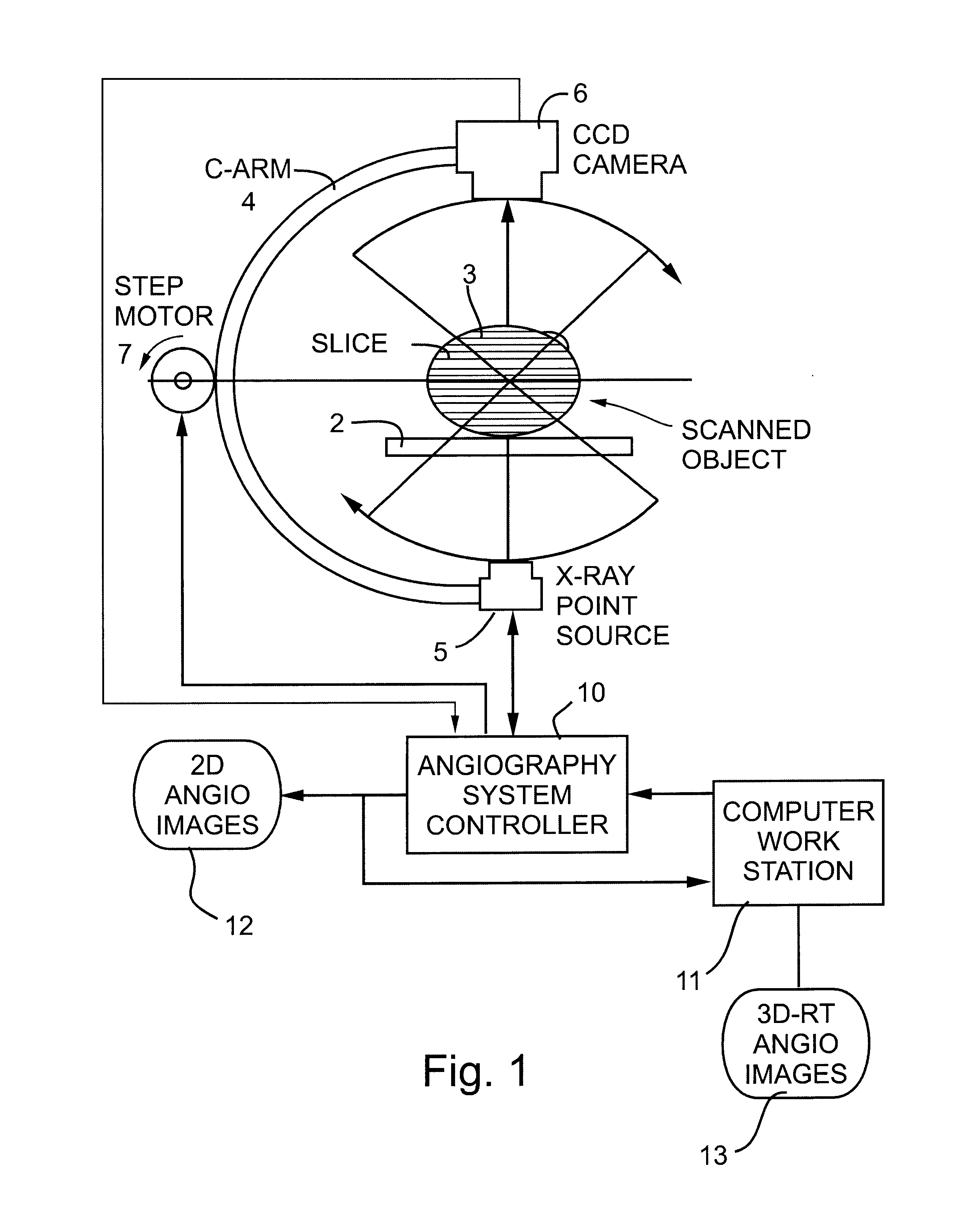
FIG. 1 schematically illustrates one form of apparatus constructed in accordance with the present invention particularly useful for producing either two-dimensional angiographs and / or three-dimensional angiographs of a patient's vascular system.

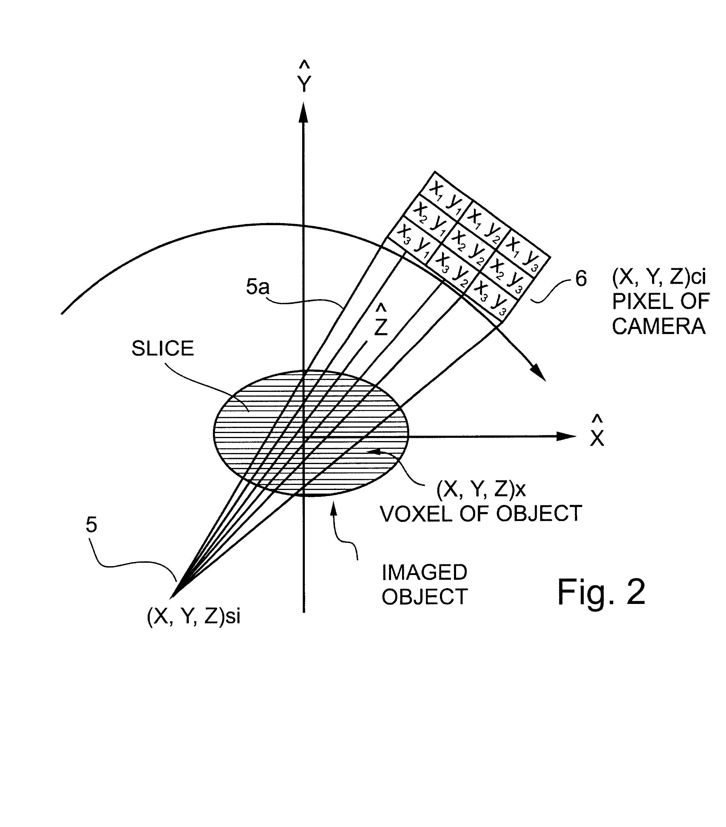
The system illustrated in FIG. 1 includes a horizontal support such as a table 2 for the patient 3 under examination, and a gantry C-arm 4, such as used in CT examination apparatus, enclosing the patient's body 3. The C-arm supports a radiation source 5 at one side of the patient's body, and a radiation detector 6 at the opposite side and in alignment with the radiation source. The radiation source 5 is an X-ray point source which produces a conical beam 5a as shown in FIG. 2; and the radiation detector 6, preferably a CCD camera, includes a two-dimensional matrix of detector elements as best seen in FIG. 4. As shown particularly in FIG. 2, the conical beam 5a produced by the radiation source 5 in each angular posit...


PUM


Abstract

A method and apparatus for angiographically imaging a portion of a patient's vascular system, by:(a) producing a first sequence of masking images of the portion of the patient's vascular system;(b) determining the masking image in the sequence having a minimum change over its immediately preceding masking image in the sequence;(c) injecting a contrast material into the patient's vascular system;(d) producing a corresponding sequence of contrast images of the portion of the patient's vascular system while containing the contrast material and(e) subtracting from each contrast image of the sequence the corresponding masking image in the sequence starting with the minimum change masking image, to thereby produce a difference image having an enhanced contrast-to-noise ratio (CNR).

Description

FIELD AND BACKGROUND OF THE INVENTIONThe present invention relates to methods and apparatus for imaging two-dimensional (2-D) planes or surfaces and / or three-dimensional (3-D) objects. The invention is particularly useful in angiography for imaging a person's vascular system in 2-D or 3-D, and is therefore described below with respect to this application.Electromagnetic radiation has long been used to produce images of internal structures of the human body for purposes of diagnosis or treatment of diseases. One technique which has gained widespread use since the 1970's is computerized tomography (CT), sometimes called computerized axial tomography (CAT), in which a narrow beam of X-rays is swept across an area of the body and is recorded by a radiation detector as a pattern of electrical impulses, while a computer is used to integrate the data for many such sweeps and to reconstruct a two-dimensional or a three-dimensional image of the examined volume. In the current CT systems, the...

Claims


Application Information

Patent Timeline
Patent Type & Authority Patents(United States)
IPC IPC(8): A61B6/00
CPCA61B6/481A61B6/504
Inventor KEREN, HANAN
Owner MEDIMAG C V I
Why Eureka
Try Eureka