System and method for enhancing confocal reflectance images of tissue specimens

a tissue specimen and reflectance imaging technology, applied in the field of confocal microscopy, can solve the problems of difficult confocal detection of cancers, and achieve the effect of increasing the contrast of nuclei and being easy to observ

Inactive Publication Date: 2006-09-19
CALIBER IMAGING & DIAGNOSTICS +1
View PDF14 Cites 7 Cited by
  • Summary
  • Abstract
  • Description
  • Claims
  • Application Information

AI Technical Summary

Benefits of technology

[0009]It has been found in accordance with the invention that a confocal laser scanning microscope using cross polarized components of light in illumination and in the detection of the reflected light from tissue specimens immersed in such an enhancement agent solution images of the cellular structure are enhanced, enabling cells and voids in the structure and the cell condition to be readily observed. By virtue of the use of such cross polarized light in imaging of tumor slices obtained in the course of Mohs surgery, epidermis sections which may have holes in the collagen are imaged more accurately so that holes are unlikely to be confused with cells or cell structure.
[0010]A method is also provided for detecting cancerous basal cell and

Problems solved by technology

Thus, when neoplastic epidermal cells invade the dermis as in BCCs and SCCs, confocal detection of the canc

Method used


Image

  • System and method for enhancing confocal reflectance images of tissue specimens
  • System and method for enhancing confocal reflectance images of tissue specimens
  • System and method for enhancing confocal reflectance images of tissue specimens

Examples


Embodiment Construction

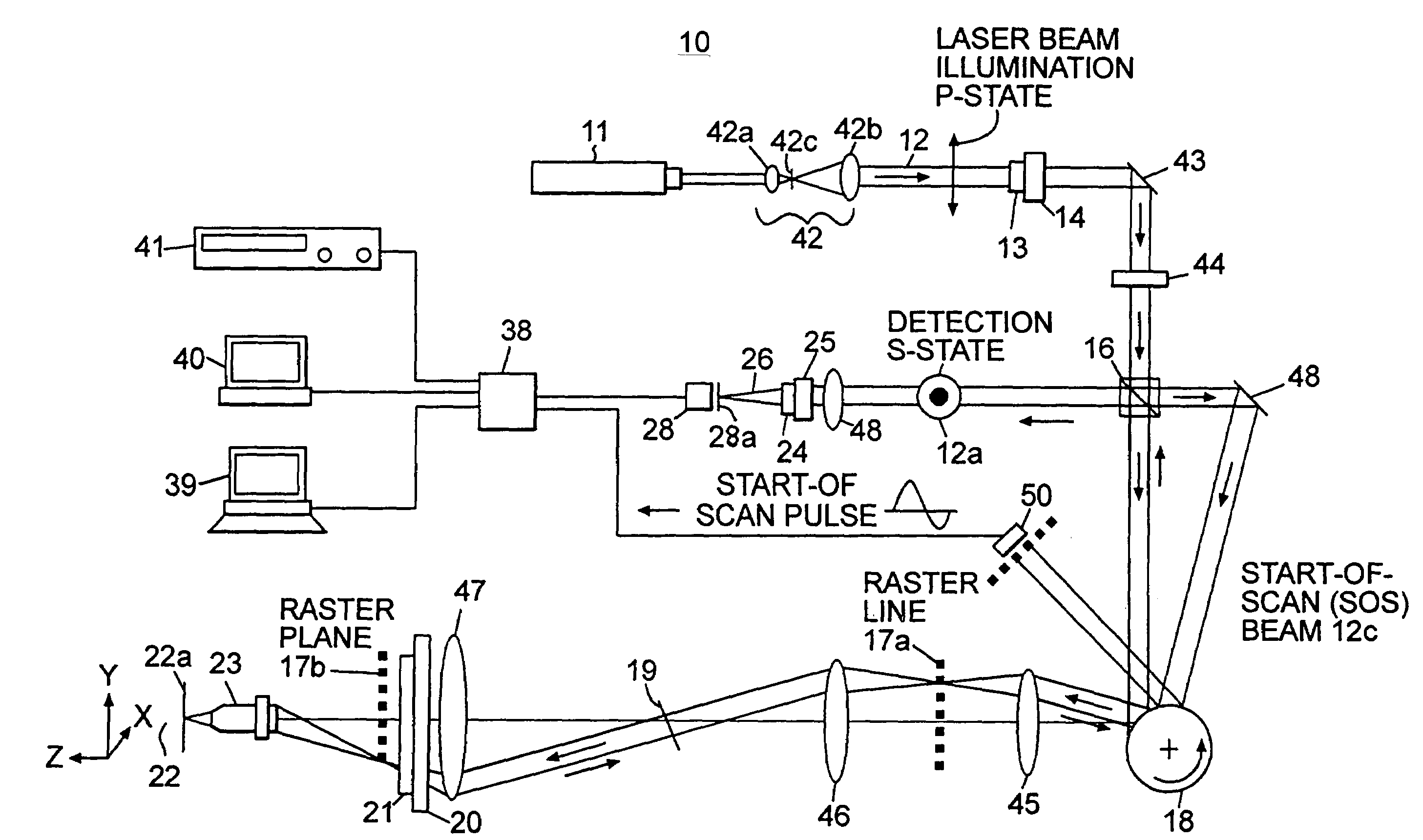
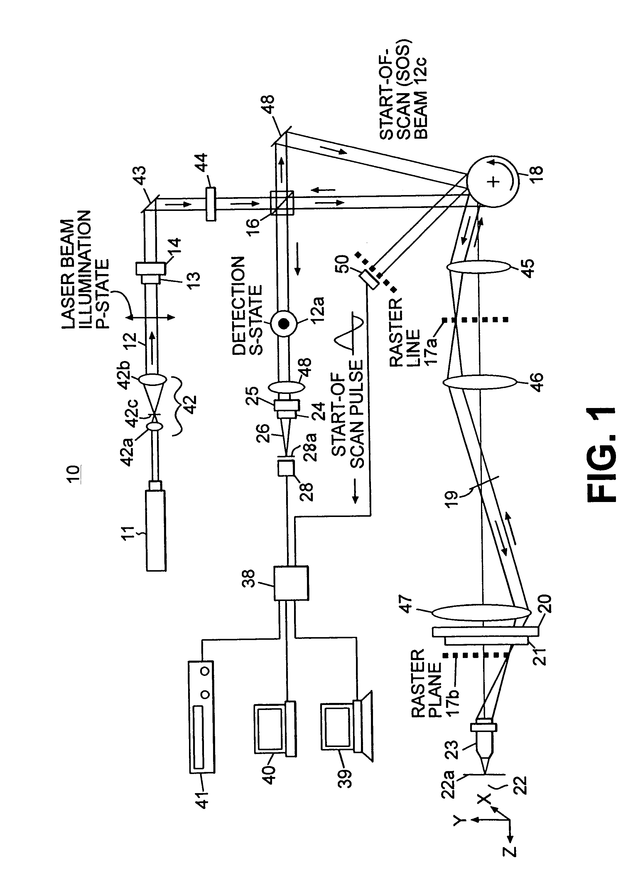
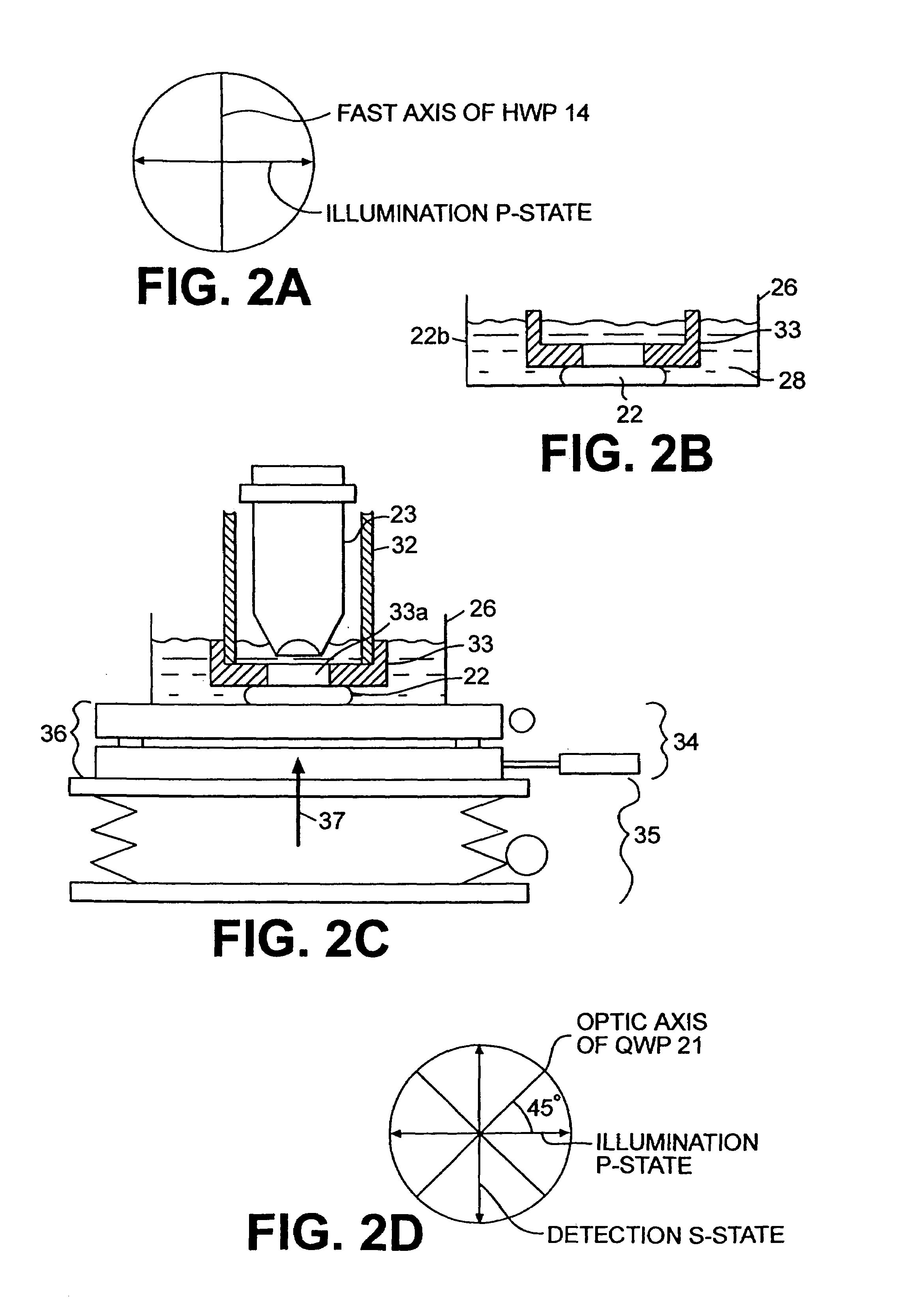
[0014]Referring to the drawings, in the confocal microscope 10 of FIG. 1, a linearly polarized (p-state) laser beam 12 is passed through a half wave plate (HWP) 13 on a rotation stage 14. A confocal microscope especially suitable in practicing the invention is described in U.S. Pat. No. 5,880,880, issued Mar. 9, 1999, which is herein incorporated by reference. Other confocal microscopes may also be used. The illumination through the non-polarizing or partially polarizing beam splitter 16 is scanned, as by a polygon mirror 18 and galvanometric mirror 19 across the specimen or sample 22 having a surface 22a. As shown in FIGS. 2B and 2C, sample 22 may be a BCC / SCC sample in a sample holder or container 22b contained in an enhancement solution bath 26 having water 28 under a tissue ring 33 which places the sample 22 under tension. As shown in FIG. 1, the microscope 10, via an objective lens 23, images the tissue sample 23 through an opening 33a in the tissue ring 33. For example, the op...


PUM


Abstract

A confocal scanning microscope system (10) using cross polarization effects and an enhancement agent (acetic acid) to enhance confocal microscope reflectance images of the nuclei of BCCs (basal cell carcinomas) and SCCs (squamous call carcinomas) in the confocal reflectance images of excised tumor slices. The confocal scanning microscope system having a laser (11) for generating an illumination beam (12), a polygon mirror (18) for scanning the beam to a tissue sample (22) and for receiving a return beam from the tissue sample and detector (28) for detecting the returned beam to form an image. The system further includes a half-waveplate (13) having a rotatable stage (14) and a quarter-wave plate (21) having a rotatable stage (20) disposed in the optical path of the illumination beam and at least a linear polarizer (24) having a rotatable stage (25) disposed in the optical pat of the returned beam from the tissue sample.

Description

[0001]This application is a Continuation of application Ser. No. 09 / 936,535, filed Sep. 14, 2001, now U.S. Pat. No. 6,720,547, a National Application under 35 U.S.C. §371 of International Pat. application Ser. No. PCT / US00 / 07008, filed Mar. 17, 2000, which claims the benefit of priority to U.S. Provisional Application Nos. 60 / 125,033, filed Mar. 18, 1999, and 60 / 146,819, filed Aug. 2, 1999, which are herein incorporated by reference.FIELD OF THE INVENTION[0002]The present invention relates to confocal microscopy and particularly to a system (method and apparatus) for enhancing images of tissue at the surface or internally of a tissue sample so as to enable rapid and accurate screening of tissue for the determination of the nuclear and cellular structure thereof. The present invention also relates to a method for diagnosing cancerous cells in skin tissue using confocal microscopy. The invention is especially suitable in providing enhanced images of the nuclei of BCC / SCC (basal cell c...

Claims


Application Information

Patent Timeline
IPC IPC(8): G01J4/00G02B21/00G02B21/14G02B21/33G02B21/36
CPCG01N21/21G01N21/47G02B21/36G02B21/0048G02B21/0068G02B21/14G02B21/33G02B21/0032G01N21/49G01N2021/4735G01N2021/4792
Inventor RAJADHYAKSHA, MILINDZAVISLAN, JAMES M.
Owner CALIBER IMAGING & DIAGNOSTICS
Why Eureka
Try Eureka