Tissue anchors and medical devices for rapid deployment of tissue anchors

a tissue anchor and rapid technology, applied in the field of medical devices, can solve the problems of requiring a significant level of skill and coordination of medical professionals, manual tying and cutting sutures can be difficult, and the difficulty of perforation closure is well documented, so as to facilitate perforation closure, simple and reliable use

Active Publication Date: 2014-02-11
COOK MEDICAL TECH LLC
View PDF414 Cites 7 Cited by
  • Summary
  • Abstract
  • Description
  • Claims
  • Application Information

AI Technical Summary

Benefits of technology

[0005]The present invention provides tissue anchors, as well as related devices and methods, for closing perforations in bodily walls. The tissue anchors are simple and reliable in use, facilitate perforation closure, and are adaptable to a variety of perforation closure situations. One embodiment of a tissue anchor, structured for engagement with a tensioning member, for closing a perforation, constructed in accordance with the teachings of the present invention, generally comprises a crossbar and a strand. The crossbar has first and second opposing ends and defines a longitudinal axis. The strand is connected to the crossbar at a location between the opposing ends. The strand has a length in the range of about 5 mm to about 50 mm extending from a distal end connected to the crossbar to a proximal end having a first connector. The strand and its first connector project away from the longitudinal axis.

Problems solved by technology

Manually tying suture strands together to close a perforation can be very complex and time consuming.
For example, a significant level of skill and coordination is required by the medical professional, especially when the perforation and sutures are difficult to access within the body, such as in endoscopic or laparoscopic procedures.
The numerous difficulties with manually tying and cutting sutures are well documented.
Unfortunately, such automatic systems are often complex and costly, difficult to use, and limited to use in certain situations.

Method used


Image

  • Tissue anchors and medical devices for rapid deployment of tissue anchors
  • Tissue anchors and medical devices for rapid deployment of tissue anchors
  • Tissue anchors and medical devices for rapid deployment of tissue anchors

Examples


Embodiment Construction

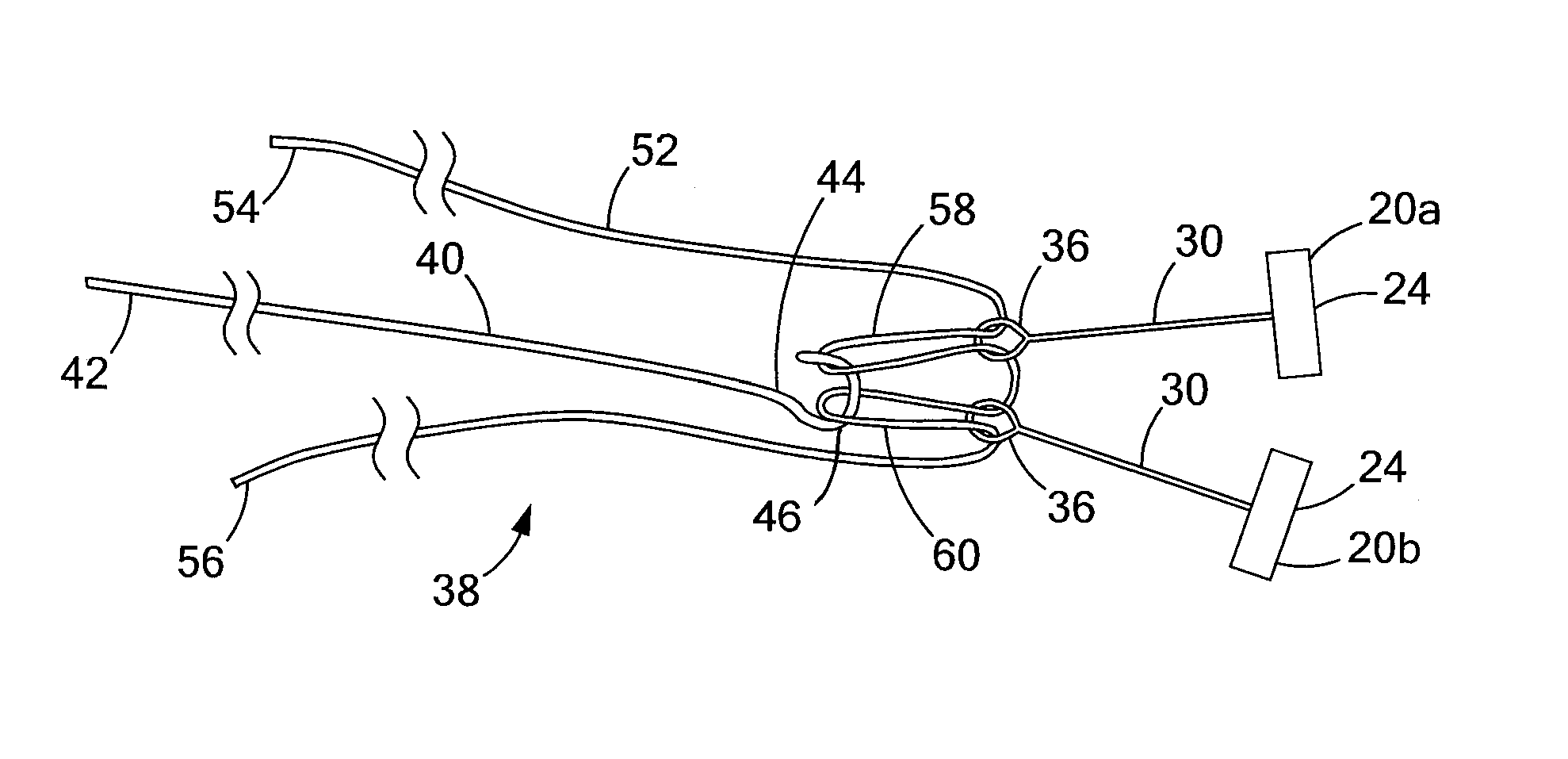
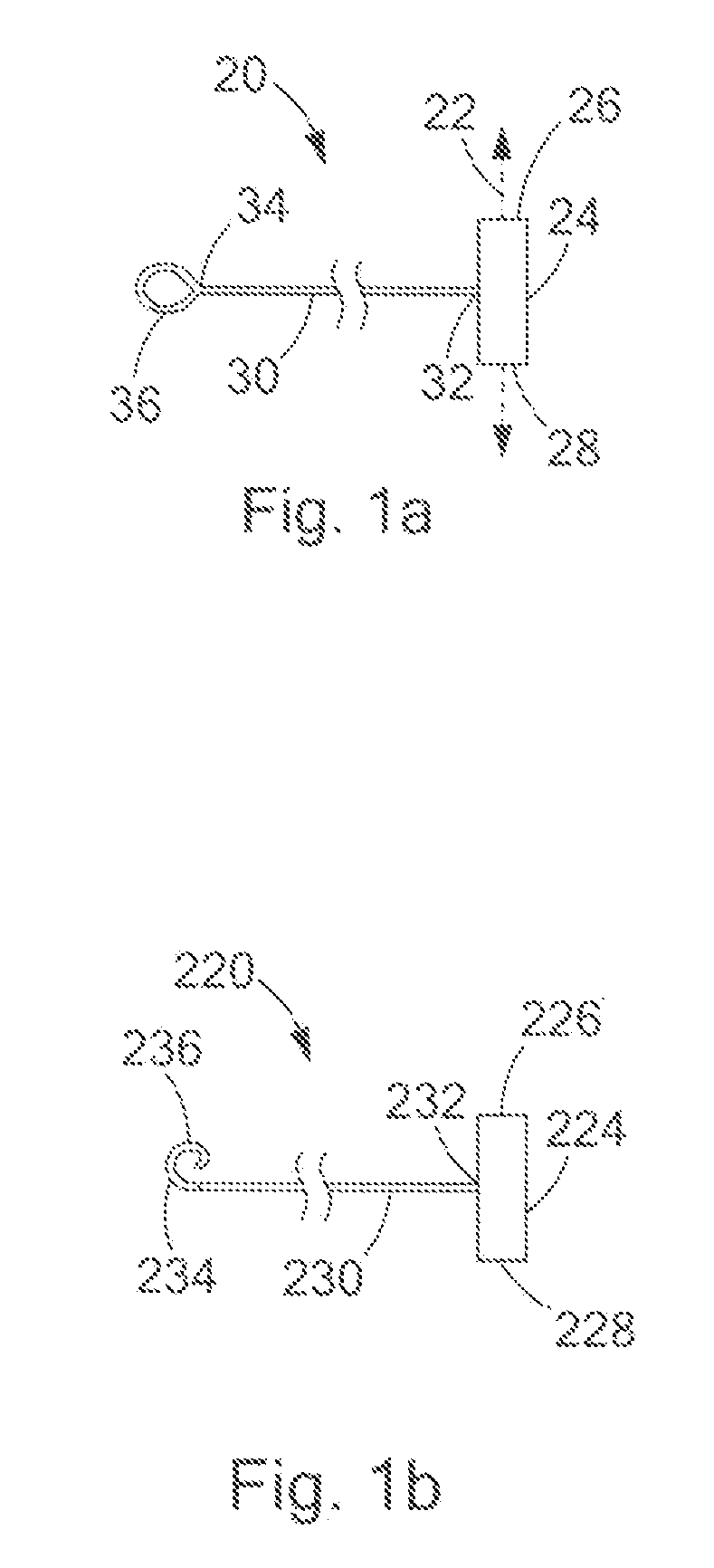
[0018]Turning now to the figures, FIG. 1a depicts a tissue anchor 20 constructed in accordance with the teachings of the present invention. The anchor 20 is utilized for closing a perforation 10 in a bodily wall 12 (FIGS. 5-7). The anchor 20 may also be used for apposing tissue, for example, in gastroesophageal reflux disease (GERD) therapy, or bariatric surgery in which an anastomosis is formed, or for use in other procedures. The anchor 20 generally includes a crossbar 24 having opposing ends 26 and 28 and defining a longitudinal axis 22. A flexible strand 30 is connected to the crossbar 24 at a location between the opposing ends 26 and 28 of the crossbar 24. The strand 30 includes a distal end 32 connected to the crossbar 24 and extending away from the longitudinal axis 22 of the crossbar 24 to a proximal end 34 which terminates in a connector 36, discussed in further detail below.

[0019]The crossbar 24 is preferably elongated, but may take any form suitable for closing the perfor...


PUM


Abstract

Medical devices and related methods are provided for closing a perforation in a bodily wall. The medical device generally includes a set of tissue anchors and an elongate tensioning member. Each anchor includes a crossbar having opposing ends and defining a longitudinal axis. A strand is connected to the crossbar at a location between the opposing ends and projects away from the longitudinal axis. The strand includes a distal end connected to the crossbar and a proximal end having a first connector. The elongate tensioning member is structured to selectively engage and disengage the first connector. The strands of the anchor are capable of being tensioned and fixed together for closing the perforation. The tensioning member is removed after the strands are fixed together.

Description

CROSS-REFERENCE TO RELATED APPLICATIONS[0001]This application claims the benefit of U.S. Provisional Application Ser. No. 61 / 166,364 filed on Apr. 3, 2009, entitled “TISSUE ANCHORS AND MEDICAL DEVICES FOR RAPID DEPLOYMENT OF TISSUE ANCHORS,” the entire contents of which are incorporated herein by reference.FIELD OF THE INVENTION[0002]The present invention relates generally to medical devices, and more particularly relates to tissue anchors for closing perforations in tissue.BACKGROUND OF THE INVENTION[0003]Perforations in bodily walls may be naturally occurring, or formed intentionally or unintentionally. In order to permanently close these perforations and allow the tissue to properly heal, numerous medical devices and methods have been developed employing sutures, adhesives, clips, staples and the like. Many of these devices typically employ one or more sutures, the strands of which must be brought together and fixed in place in order to close the perforation, and thereafter cut a...

Claims


Application Information

Patent Timeline
Patent Type & Authority Patents(United States)
IPC IPC(8): A61B17/04
CPCA61B2017/0417A61B17/0057A61B17/0401A61B2017/00663
Inventor DUCHARME, RICHARD, W.
Owner COOK MEDICAL TECH LLC
Why Eureka
Try Eureka