Diagnostic method for brain damage-related disorders
a brain damage and diagnostic method technology, applied in the direction of biological material analysis, peptides, drug compositions, etc., can solve the problems of poor sensitivity and specificity, hampered diagnostic value of brain damage-related biomarkers, and inability to accurately predict clinical status and functional outcomes
- Summary
- Abstract
- Description
- Claims
- Application Information
AI Technical Summary
Benefits of technology
Problems solved by technology
Method used
Image

Examples
example 1
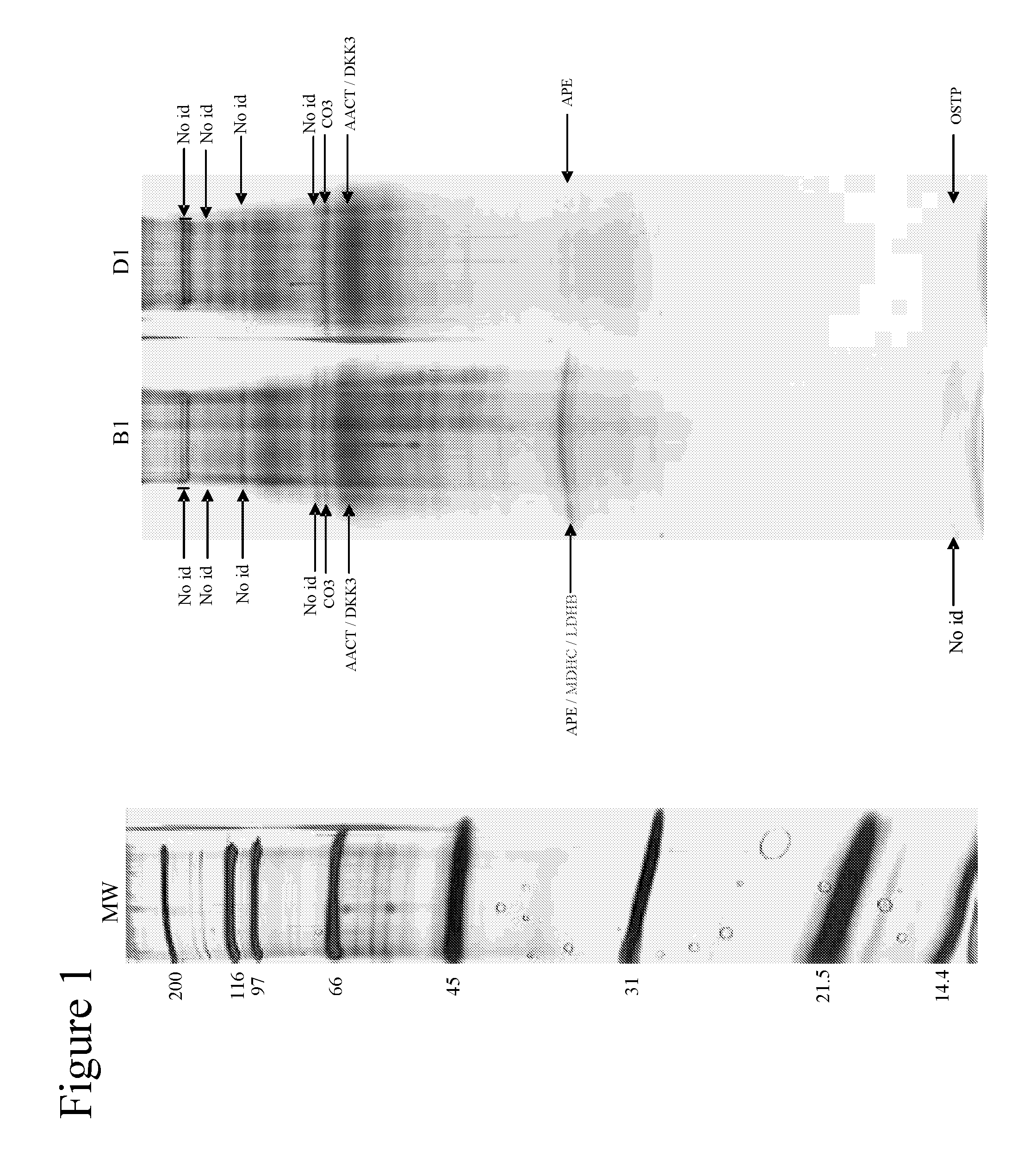
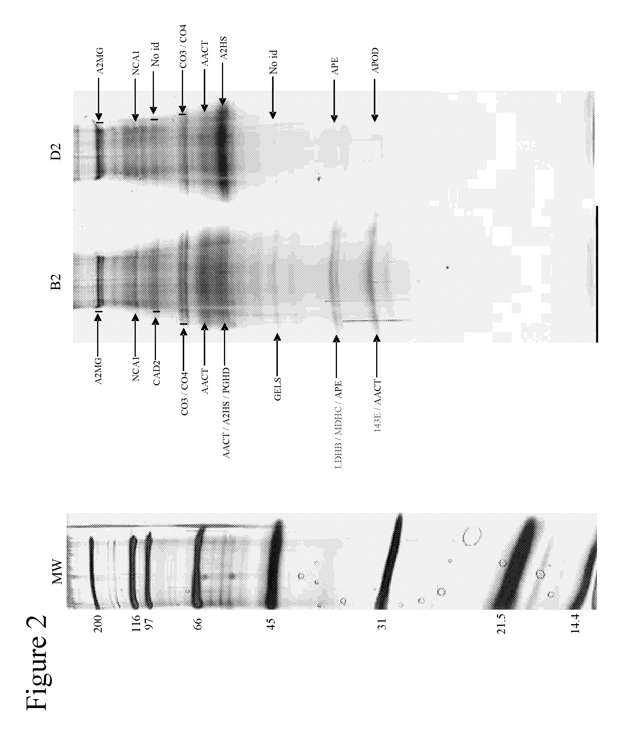
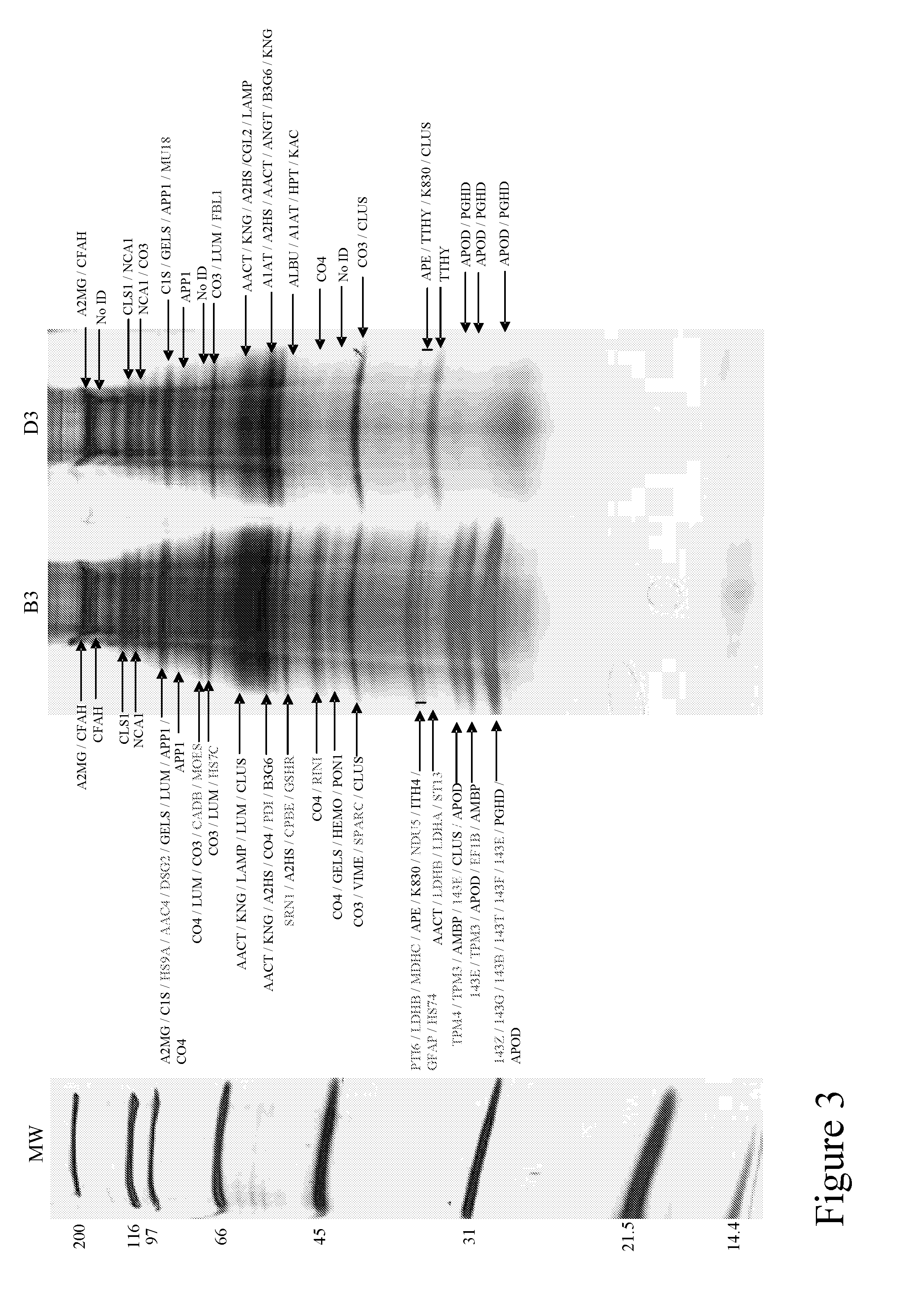
[0088]Using one-dimensional gel electrophoresis (1-DE) separation of cerebrospinal fluid (CSF) proteins and mass spectrometry techniques, 58 polypeptides named in Table 1 were found elevated or decreased in the CSF of deceased patients, used as a model of massive brain damage.
Study Population and Sample Handling
[0089]Twenty CSF samples were used for the proteomics-based approach aiming at discovering brain damage-related disorder markers. Five of these samples were obtained at autopsy from deceased patients 6 hours after death with no pathology of the central nervous system. Fifteen others were collected by lumbar puncture from living patients who had a neurological workup for benign conditions unrelated to brain damage (atypical headache and idiopathic peripheral facial nerve palsy). CSF samples were centrifuged immediately after collection, aliquoted, frozen at −80° C. and stored until analysis.
CSF Depletion Fractionation
[0090]Immunodepletion of human serum albumin, transferrin, h...
example 2
Introduction
[0094]One of the proteins identified as being upregulated in deceased CSF was evaluated as a potential biomarker of cerebrovascular disease, an example of a brain damage-related disorder. A survey of stroke patients was carried out and the results are shown in FIGS. 5 to 7. An ELISA intensity signal was obtained for Ubiquitin fusion degradation protein 1 homolog (UFD1) in plasma samples of the patients and of negative control patients. Plasma samples were taken from patients between 0-24 hours and / or after 72 hours of arrival at emergency hospital, and were matched for age / sex with samples from control patients.
[0095]ELISA was performed using 96-well Reacti-Bind™ NeutrAvidin™ coated Black Plates (Pierce, Rockford, Ill.). Plates were first rinsed in Borate Buffer Saline pH 8.4 (BBS) (100 mM H3BO3, 25 mM Na2B4O7 (Sigma, St Louis, Mo., USA), 75 mM NaCl (Merck, Darmastadt, Germany)) on a NOVAPATH™ washer (Bio-Rad, Hercules, Calif.). Then, 50 μl of biotin-conjugated antibody ...
example 3
[0099]This Example provides additional data showing plasma levels of UFDP1 in stroke and control patients. Additional data has been obtained from two cohorts of patients and controls, the smaller from Geneva, and a more comprehensive panel from the US.
[0100]ELISA was performed using 96-well Reacti-Bind™ NeutrAvidin™ coated Black Plates (Pierce, Rockford, Ill.). Plates were first rinsed in Borate Buffer Saline pH 8.4 (BBS) (100 mM H3BO3, 25 mM Na2B4O7 (Sigma, St Louis, Mo., USA), 75 mM NaCl (Merck, Darmastadt, Germany)) on a NOVAPATH™ washer (Bio-Rad, Hercules, Calif.). Then, 50 μl of relevant biomarker specific biotin-conjugated antibody (2 μg / mL) prepared in the dilution buffer A at pH 7 was added and incubated for one hour at 37° C. Plates were then washed 3 times in BBS in the plate washer. 50 μl of antigen or plasma was then added and incubated for one hour at 37° C. Recombinant protein antigens were diluted at 100, 50, 25, 12.5, 6.25, 3.125, 1.56 ng / ml in dilution buffer A to g...
PUM
| Property | Measurement | Unit |
|---|---|---|
| concentrations | aaaaa | aaaaa |
| concentrations | aaaaa | aaaaa |
| temperature | aaaaa | aaaaa |
Abstract
Description
Claims
Application Information
 - R&D
- Intellectual Property
- Life Sciences
- Materials
- Tech Scout
- Unparalleled Data Quality
- Higher Quality Content
- 60% Fewer Hallucinations
Browse by: Latest US Patents, China's latest patents, Technical Efficacy Thesaurus, Application Domain, Technology Topic, Popular Technical Reports.
© 2025 PatSnap. All rights reserved.Legal|Privacy policy|Modern Slavery Act Transparency Statement|Sitemap|About US| Contact US: help@patsnap.com