
Acellular matrix implanted into an articular cartilage or osteochondral lesion protected with a biodegradable polymer modified to have extended polymerization time and methods for preparation and use thereof
Patent Information
- Authority / Receiving Office
- US · United States
- Current Assignee / Owner
- Publication Date
- 2005-02-24
- Estimated Expiration
- Not applicable · inactive patent
Smart Images
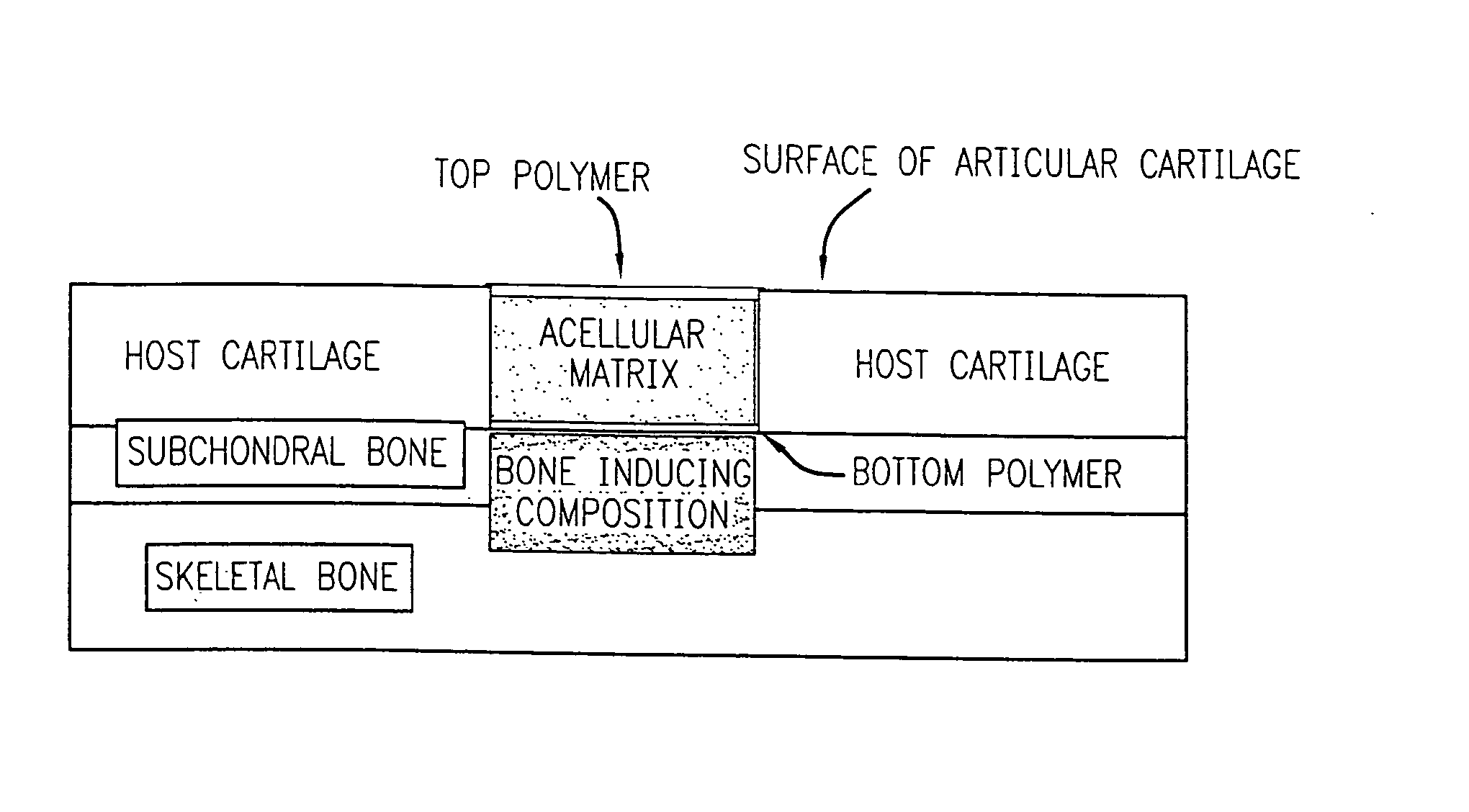
Figure 1 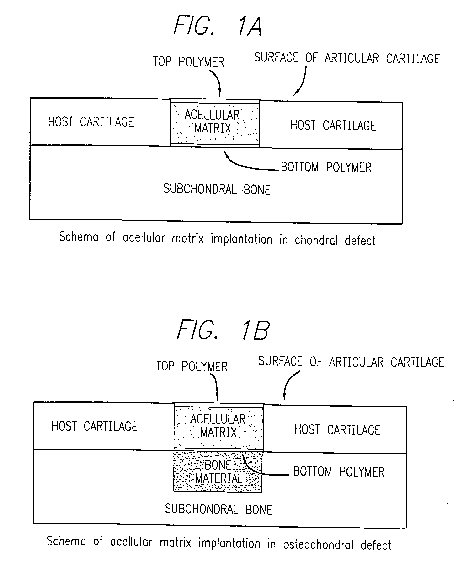
Figure 2 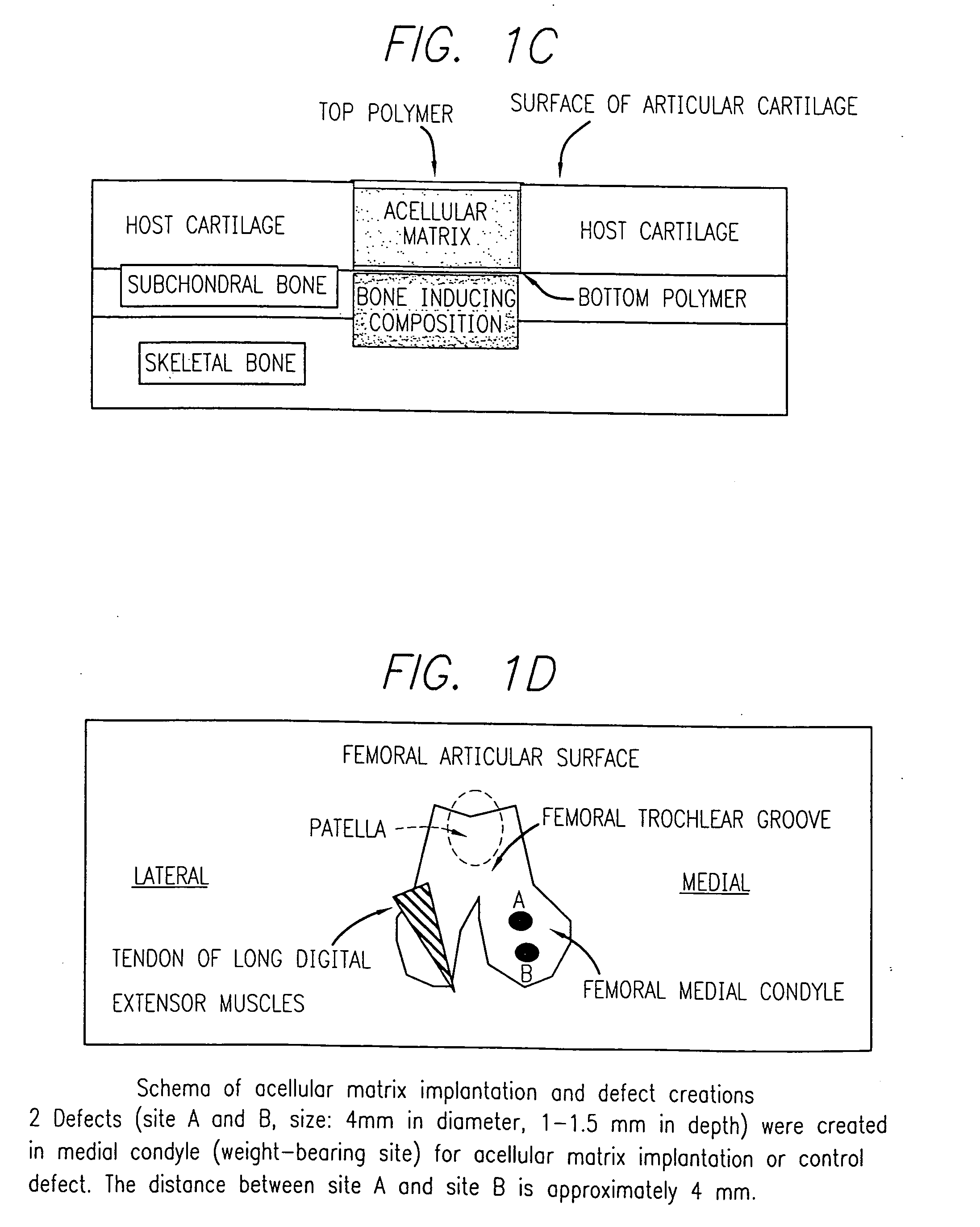
Figure 3
Abstract
Description
BACKGROUND OF THE INVENTION This application is based on and claims priority of the Provisional Application Serial No.: 60 / 496,971, filed Aug. 20, 2003. FIELD OF INVENTION The current invention concerns acellular matrix implants implanted into an articular cartilage or osteochondral lesion protected with a biodegradable polymer barrier modified to have extended polymerization time. The invention further concerns compositions for treatment of articular cartilage or osteochondral defects and injuries and a method for treatment of articular cartilage or osteochondral defects using an acellular matrix implant implanted into a joint cartilage lesion and / or into the osteochondral defect in situ wherein the osteochondral defect is further implanted with a bone-inducing composition or a carrier comprising said composition. In particular, the current invention concerns acellular matrix implants implanted into the articular cartilage or osteochondral lesion protected with a protective biode...
Examples
example 1
Preparation of Acellular Collagenous Implants
This example illustrates preparation of the acellular matrix implant.
300 grams of a 1% aqueous atelocollagen solution (VITROGEN®), maintained at pH 3.0, is poured into a 10×20 cm tray. This tray is then placed in a 5 liter container. A 50 ml open container containing 30 ml of a 3% aqueous ammonia solution is then placed next to the tray, in the 5 liter chamber, containing 300 grams of said 1% aqueous solution of atelocollagen. The 5 liter container containing the open trays of atelocollagen and ammonia is then sealed and left to stand at room temperature for 12 hours. During this period the ammonia gas, released from the open container of aqueous ammonia and confined within the sealed 5 liter container, is reacted with the aqueous atelocollagen resulting in gelling said aqueous solution of atelocollagen.
The collagenous gel is then washed with water overnight and, subsequently, freeze-dried to yield a sponge like matrix. This freeze ...
example 2
Biochemical and Histological Assays
This example describes assays used for biochemical and histological studies.
For biochemical (DMB) assay, the implant taken from the animal after certain time following the implantation, transferred to microcentrifuge tubes and digested in 300 μl of papain (125 μg / ml in 0.1 M sodium phosphate, 5 mM disodium EDTA, and 5 mM L-cysteine-HCl) for 18 hours at 60° C. S-GAG production in the implant is measured using a modified dimethylene blue (DMB) microassay with shark chondroitin sulfate as a control according to Connective Tissue Research, 9: 247-248 (1982).
DNA content is determined by Hoechst 33258 dye method according to Anal. Biochem., 174:168-176 (1988).
For histological assay, the remaining implants from each group were fixed in 4% paraformaldehyde. The implants were processed and embedded in paraffin. 10 μm sections were cut on a microtome and stained with Safranin-O (Saf O).
For immunohistochemistry, the samples are contacted with diamin...
example 3
Evaluation of Integration of Acellular Matrix Implant in a Swine Model
This example describe the procedure and results of study performed for evaluation of integration of porcine in a swine model.
An open arthrotomy of the right knee joint was performed on all animals, and a biopsy of the cartilage was obtained.
A defect was created in the medial femoral condyle of the pig,s right knee. This defect (control) was not implanted with an acellular matrix implant but was left intact. Following surgery, the joint was immobilized with an external fixation implant for a period of about two weeks. Two weeks after the arthrotomy on the right knee was performed, an open arthrotomy was performed on the left knee and defects were created in this medial femoral condyle. The acellular matrix implant was implanted within the defect (s) in this knee which was similarly immobilized. The operated sites were subsequently viewed via arthroscopy two weeks after implantation or defect creation and ther...