X-ray diagnostic imaging apparatus and x-ray apparatus

An image diagnosis and X-ray technology, which is applied in the fields of radiological diagnosis instruments, diagnosis, medical science, etc., and can solve problems such as redundant space

Active Publication Date: 2012-12-12
TOSHIBA MEDICAL SYST CORP
View PDF3 Cites 0 Cited by
  • Summary
  • Abstract
  • Description
  • Claims
  • Application Information

AI Technical Summary

Problems solved by technology

Therefore, for the examination room, it is necessary to have an extra space that is not necessarily necessary for treatment or surgery.

Method used


Image

  • X-ray diagnostic imaging apparatus and x-ray apparatus
  • X-ray diagnostic imaging apparatus and x-ray apparatus
  • X-ray diagnostic imaging apparatus and x-ray apparatus

Examples


Embodiment Construction

[0018] Several embodiments of the present invention are described below with reference to the accompanying drawings. In the drawings, the same reference numerals denote the same parts.

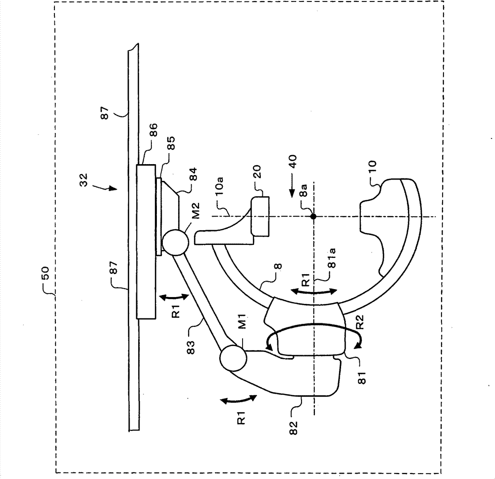
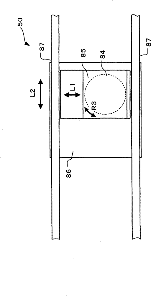
[0019] Below, refer to Figure 1 to Figure 7 An X-ray image diagnostic apparatus according to a first embodiment of the present invention will be described.

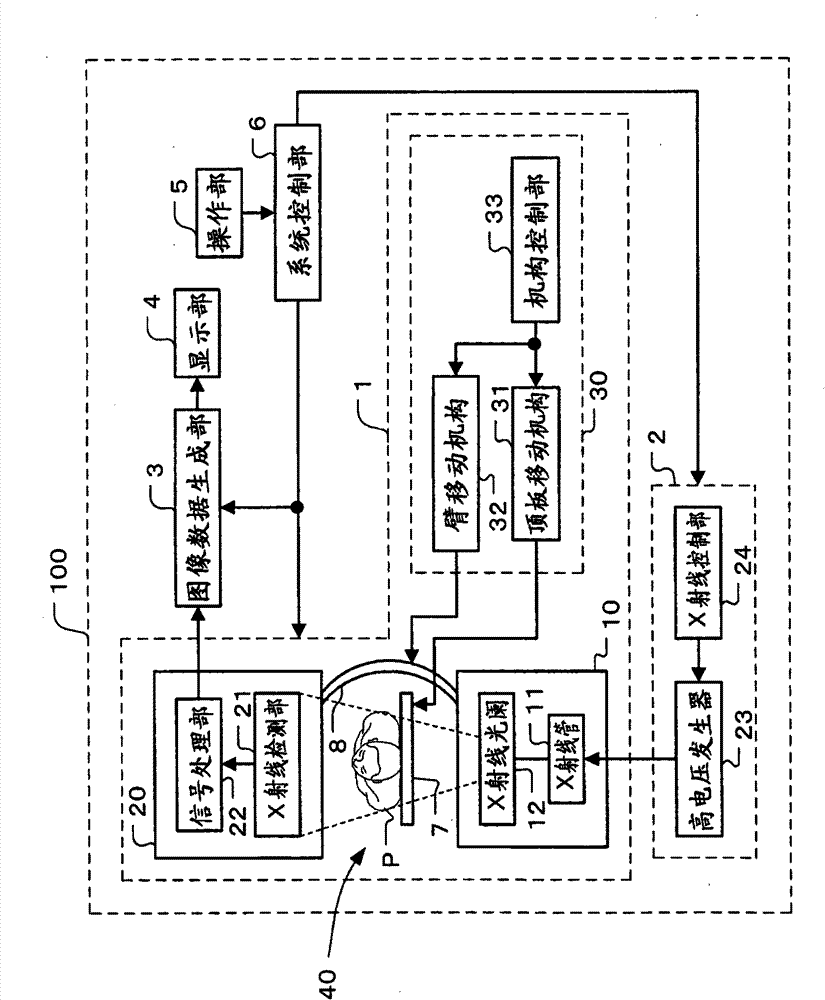
[0020] figure 1 is a block diagram showing the configuration of the X-ray image diagnostic apparatus according to the first embodiment of the present invention.

[0021] like figure 1 As shown, the X-ray image diagnostic apparatus 100 includes: X-ray irradiation / detection part (component) 1, high voltage generation part (unit) 2, image data generation part (unit) 3, display part 4, operation part 5 and system control Part 6.

[0022] The X-ray irradiation / detection unit (means) 1 performs X-ray imaging of the subject P. As shown in FIG. The high voltage generating unit (unit) 2 generates a high voltage necessary for X-ray imaging o...


PUM


Abstract

An X-ray generation unit and an X-ray detection unit are held by an arm. Image data is generated based on X-ray projection data which is generated by the X-ray detection unit. A first support member supports the arm and rotates the arm in a first rotation direction. A second support member supports the first support member. The second support member rotates the first support member around a rotation center in a second rotation direction perpendicular to the first rotation direction. A movement unit can move the second support member in an up and down direction.

Description

technical field [0001] The present invention relates to an X-ray image diagnosis device and an X-ray device used for diagnosing a subject. Background technique [0002] In recent years, with the development of catheter-based angiography and IVR (Interventional Radiology), X-ray image diagnostic devices have been progressing mainly in the field of circulatory organs. This X-ray image diagnostic device is used for percutaneous angioplasty and congenital heart malformation in combination with cardiac surgery. X-ray image diagnostic equipment, as a highly advanced IVR system, is expanding its range of applications. The environment where X-ray image diagnostic equipment is installed is also developing from a general cath lab to a composite system called a hybrid system that integrates with an operating room. [0003] Japanese Patent Laid-Open Publication No. 2008-190 discloses an X-ray image diagnostic device for circulatory organs. Such an X-ray image diagnostic apparatus is ...

Claims


Application Information

Patent Timeline
Patent Type & Authority Patents(China)
IPC IPC(8): A61B6/00
CPCA61B6/4441A61B6/587A61B6/4458A61B6/4464
Inventor 野田浩二
Owner TOSHIBA MEDICAL SYST CORP
Why Eureka
Try Eureka