Method for identifying cancer cells

An identification method and technology for cancer cells, applied in the field of cancer cell identification, can solve the problems of prone to errors, increased detection complexity, and high price.

Inactive Publication Date: 2011-09-07
樊潮
View PDF5 Cites 19 Cited by
  • Summary
  • Abstract
  • Description
  • Claims
  • Application Information

AI Technical Summary

Problems solved by technology

[0004] (1) Cancerous cells have no definite shape: the variation of nucleus and cell membrane is irregular;
[0005] (2) Cell identification under multi-angle cannot be realized: mutated cells exist at any angle, and the detection complexity increases;
[0007] (4) The complexity of detection is related to medical conditions, and manual detection cannot complete the detection of massive image data;
[0018] (10) Pathological examination: including cytology and biopsy tissue examination. The final diagnosis of cancer must have a pathological certificate. The advantage of pathological examination is that it is accurate, but the disadvantage is that the workload is heavy and errors are prone to occur;
[0019] The above detection methods (1)-(9) are not only expensive, ranging from thousands of yuan to tens of thousands of yuan. For example, CT detection, although the effect is very good, but each detection is about 10,000 yuan, which makes many Ordinary people are discouraged, and can't make analysis at the cell level. In many cases, when a tumor is detected, the patient is declared to be in the middle or late stage, which is not an early detection in the true sense.

Method used


Image

  • Method for identifying cancer cells
  • Method for identifying cancer cells
  • Method for identifying cancer cells

Examples


Embodiment Construction

[0110] The method of the present invention will be described in further detail below in conjunction with the accompanying drawings and embodiments.

[0111] A cancer cell identification method of the present invention comprises the following steps:

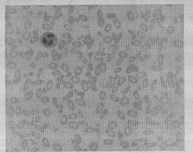
[0112] Step 1: Acquisition of images: First, the specimen is prepared, and then images are acquired through a Carl Zeiss microscope such as figure 1 As shown, the minimum configuration of the microscope to the computer is: Pentium 4 1.8GHZ, 512KB memory, 80GB hard disk; support 1280×1024 resolution graphics card and support 32-bit color depth; support IEEE 1394 interface, the process schematic diagram of this embodiment is as follows figure 2 shown;

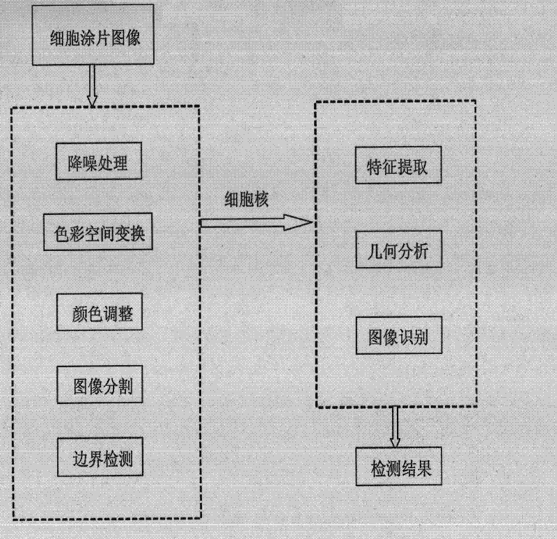
[0113] Step 2: Process the collected images, as shown in the schematic diagram image 3 As shown, the process is as follows:

[0114]Establish a sample image library of normal human cells, and analyze these images to study the distribution of normal human cells, the method is as...


PUM


Abstract

The invention relates to a method for identifying cancer cells, belonging to the field of a medical instrument. The method comprises the following steps of: collecting images, analyzing the images, and outputting detection results, wherein the image analysis comprises noise reduction treatment, color space exchange, color regulation, images cutting, characteristic extraction, geometric analysis, and image identification. The invention provides a system and method for identifying cancer cells disclosed by the invention have reasonable price and good detection effect, can automatically analyze pathological images so as to find tumor cells at the early stage.

Description

technical field [0001] The invention belongs to the field of medical devices, in particular to a method for identifying cancer cells. Background technique [0002] Cancer has become the number one killer that threatens human health in modern society, but as we all know, if early detection, early diagnosis, and early treatment can be achieved, the cure rate of cancer patients can be improved, and the mortality rate can be delayed and reduced. Diagnosis mainly refers to when the tumor is still in the initial growth stage, the diseased tissue is still limited to a small part of the normal tissue, and the patient is found and diagnosed when the patient has no obvious symptoms or only mild symptoms. Early detection is very important for the treatment and overcoming of cancer. It is of vital significance. According to statistical data, among the cancer patients seen in the outpatient departments of hospitals in large and medium-sized cities, early-stage patients account for about ...

Claims


Application Information

Patent Timeline
Patent Type & Authority Applications(China)
IPC IPC(8): G01N21/27C12Q1/02G06T7/00
Inventor 樊潮李建华候赛因付志民宫伟李伟
Owner 樊潮
Why Eureka
Try Eureka