
Method for culturing autologous peripheral blood lymphocytes
A technology of lymphocytes and peripheral blood, which is applied in the field of in vitro culture of immune cells, can solve the problems of low expansion efficiency of autologous lymphocyte culture technology, great impact on the patient's body, and weak tumor killing activity of cells, so as to achieve activation and expansion efficiency Improvement, high activity and stability, and the effect of improving activation efficiency and amplification efficiency
- Summary
- Abstract
- Description
- Claims
- Application Information
AI Technical Summary
Problems solved by technology
Method used
Image

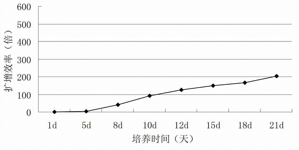
Examples
Embodiment 1
[0072] A method for cultivating autologous peripheral blood lymphocytes, comprising the steps of:
[0073] (1) After separating PBMC from 50-100mL peripheral blood, resuspend in 50mL serum-free medium, the cell concentration of PBMC is 2×10 6 cells / mL, placed at 37°C, 7.5% CO by volume 2 , 100% relative humidity of CO 2 Static culture in the incubator for 72 hours to obtain the primary culture solution;
[0074] (2) Add serum-free medium to 200 mL of the initial culture medium prepared in step (1), and add IL-2 at the same time, so that the concentration of IL-2 is 1×10 3 U / mL, placed at 37°C, 7.5% CO by volume 2 , 100% relative humidity of CO 2 Stand and cultivate in the incubator for 96 hours to obtain the secondary culture solution;
[0075] (3) Transfer the secondary culture solution prepared in step (2) to a 1.8L cell culture bag, add serum-free medium to 1000mL, and add IL-2 at the same time, so that the concentration of IL-2 is 1×10 3 U / mL, placed at 37°C, 7.5% CO...
Embodiment 2
[0093] The method described in Example 1, the difference is that every milliliter of serum-free medium contains the following components:
[0094] IFN-γ 1500U, PHA-p 750ng, IL-1α 150U, CD3mAb 75ng, CD28mAb 75ng.
[0095] Experimental results
[0096] The detection process is as described in Example 1. After testing,
[0097] 1. Determination of cell proliferation multiple
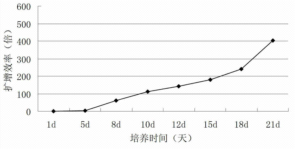
[0098] The results showed that the expansion factor of AIL cells gradually increased over time from 1 to 21 days of culture, basically showed a linear trend from 1 to 5 days, and the expansion efficiency remained constant. The reasons for the amplification factor basically showed a linear increase, and the amplification efficiency increased on the 18th to 21st day, and reached the maximum value on the 21st day of cultivation. Compared with Example 1, the maximum amplification factor was increased by 1 times, and the results were as follows: figure 2 shown.
[0099] 2. Cytotoxicity experiment
[0100]...
Embodiment 3
[0104] The method described in Example 1, the difference is that every milliliter of serum-free medium contains the following components:
[0105] IFN-γ 2000U, PHA-p 1000ng, IL-1α 200U, CD3mAb 100ng, CD28mAb 100ng.
[0106] Experimental results
[0107] The detection process is as described in Example 1. After testing,
[0108] 1. Determination of cell proliferation multiple
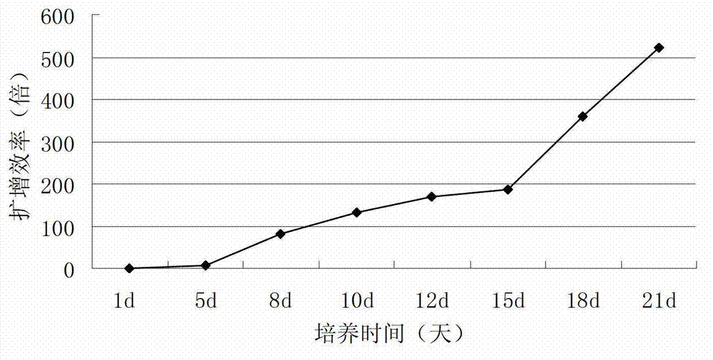
[0109] The results showed that the amplification factor of AIL gradually increased over time from 1 to 21 days of culture, basically showed a linear trend on 1 to 5 days, and the expansion efficiency remained constant. The reason is that the amplification factor basically rises linearly, and the amplification efficiency increases linearly from 18 to 21 days, and reaches the maximum value on the 21st day of cultivation. Compared with Example 2, the maximum amplification factor increases by about 1 / 3, The result is as image 3 shown.
[0110] 2. Cytotoxicity experiment
[0111] The results showed th...
PUM
 Abstract
Description
Claims
Application Information