CT-XA-image- multi-dimensional fused-based simulation navigation system and method

A CT-XA and CT image technology, applied in the field of medical device data image processing, can solve the problems of manual completion, high integrity requirements for blood vessel extraction, and inability to achieve CT image and DSA image fusion, so as to increase the amount of information.

Inactive Publication Date: 2014-07-02
CREALIFE MEDICAL TECH BEIJING
View PDF4 Cites 17 Cited by
  • Summary
  • Abstract
  • Description
  • Claims
  • Application Information

AI Technical Summary

Problems solved by technology

2D-3D registration is a complex space transformation process. It is difficult for doctors to complete this process manually. It is impossible to simulate the posture and shape of the catheter head, and it is impossible to realize CT equipment to simulate DSA catheter inspection images and realize CT images and DSA images. fusion etc.
For the point set registration algorithm, the biggest problem with this type of algorithm is that it has high requirements on the integrity of blood vessel extraction, so XA blood vessel segmentation often needs to be done manually, which is a very heavy operation for doctors.

Method used


Image

  • CT-XA-image- multi-dimensional fused-based simulation navigation system and method
  • CT-XA-image- multi-dimensional fused-based simulation navigation system and method
  • CT-XA-image- multi-dimensional fused-based simulation navigation system and method

Examples


Embodiment Construction

[0070] The technical scheme of the present invention will be described in detail below in conjunction with the examples of the present invention

[0071] 1. XA image blood vessel extraction

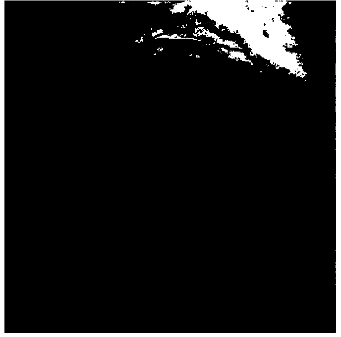
[0072] Select a frame of image in the XA image, and record the main angle and sub-angle information in the DICOM information of the image, as well as the position information of the transmitting source and receiving board. Using multi-scale Hessian convolution pairs figure 1 Perform enhancement, and count the optimal enhancement effect at all scales. The calculation formula at each scale is

[0073] V ( s ) = 0 if λ 2 > 0 , exp ( ...


PUM


Abstract

The invention discloses a CT-XA-image-multi-dimension-fusion-based-based simulation navigation system and method. A CT simulated XA image and a CT-XA fused image can be obtained through a CT coronary artery 3D image extraction and rebuilding module, a CT coronary artery analysis module, a CT coronary artery 3D image simulation XA angiocardiography 2D image module and a CT coronary artery 3D image and XA angiocardiography 2D image registration fusing and displaying module; the high registration accuracy and the whole process full automation can be achieved without accurately dividing an XA blood vessel, a plurality of common XA angiocardiography positions are preset, and angiocardiography position user defining can be further supported. By means of the CT-XA-image-multi-dimension-fusion-based-based simulation navigation system and method, the problem that the XA blood vessel cannot be displayed due to blood vessel blocking is well solved, the position and the direction of the blood vessel are further obtained, and the multi-position and multi-step single-frame single-branch blood vessel fusing navigation of XAs and the CT coronary artery is achieved.

Description

technical field [0001] The invention relates to medical device data image processing, in particular to a simulation navigation system and method based on CT-XA image multi-dimensional fusion. Background technique [0002] Using CT (Computer X-ray tomography technique, computer X-ray tomography technology) to perform enhanced scans on the heart, a complete CT image of the heart at a certain phase can be obtained. XA (X-ray angiography, X angiography), the degree of development is much better. As far as the current technology is concerned, the cardiovascular (hereinafter referred to as coronary artery) can be completely extracted from the heart image of CT enhanced scan. The same vessel with occlusion is very difficult to be visualized by XA angiography, but it can be visualized relatively easily by enhanced CT scan and extracted by technical means. In this way, the XA image can be approximated by performing projection transformation on the three-dimensional coronary image (...

Claims


Application Information

Patent Timeline
Patent Type & Authority Applications(China)
IPC IPC(8): A61B6/03A61B6/00A61B19/00
Inventor 王民汉周宇李晓光
Owner CREALIFE MEDICAL TECH BEIJING
Why Eureka
Try Eureka