Biomarker of perimuscular cells of testicular tubes and application of biomarker

A biomarker and cell technology, applied in animal cells, biological testing, vertebrate cells, etc., can solve the problems of slow research progress, failure to successfully separate Leydig cells and peritubular muscle-like cells, and effective separation of Leydig cells, etc. Simple operation and low cost effect

Pending Publication Date: 2022-04-12
THE FIFTH AFFILIATED HOSPITAL SUN YAT SEN UNIV
View PDF1 Cites 0 Cited by
  • Summary
  • Abstract
  • Description
  • Claims
  • Application Information

AI Technical Summary

Problems solved by technology

[0004] Due to the inability to effectively separate Leydig cells and testicular myoid cells in vitro, the research on Leydig cells and testicular myoid cells has been slow
[0005] In recent years, it has been reported that Nerve Growth Factor Receptor (NGFR) can be used as a marker for sorting Leydig cells. However, the applicant r

Method used


Image

  • Biomarker of perimuscular cells of testicular tubes and application of biomarker
  • Biomarker of perimuscular cells of testicular tubes and application of biomarker
  • Biomarker of perimuscular cells of testicular tubes and application of biomarker

Examples


Embodiment 1

[0064] The invention provides a test kit for identifying peritube myocytes, said kit should include: anti-NGFR protein antibody (Abcam, Ab8874), anti-ITGA9 protein antibody (R&D, AF3827), donkey anti-rabbit IgG secondary antibody (Donkey anti-Rabbit IgG (H+L) Highly Cross-Adsorbed Secondary Antibody, Alexa FluorPlus 488, Invitrogen, A32790), donkey anti-goat IgG secondary antibody (Donkey anti-Goat IgG (H+L) Highly Cross-Adsorbed Secondary Antibody, Alexa Fluor Plus594, Invitrogen, A32758) and PBS solution containing 5% bovine serum albumin (BSA).

Embodiment 2

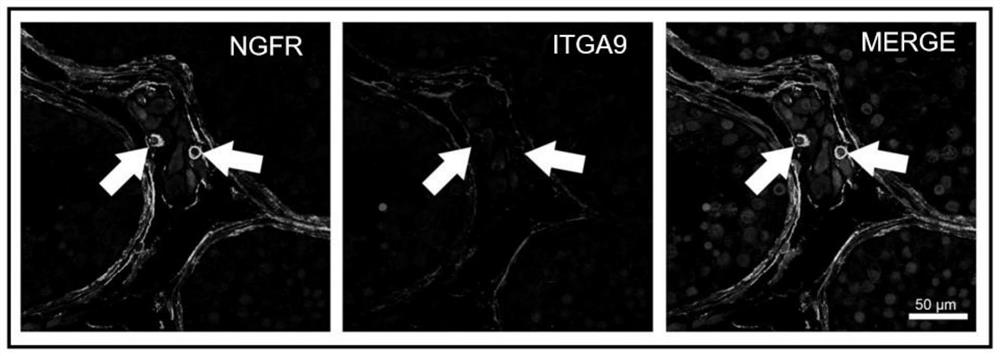
[0066] In this embodiment, immunofluorescence staining of NGFR and ITGA9 proteins in human testis tissue comprises the following steps:

[0067] 1) Tissue paraffin embedding and sectioning: The fresh testicular tissue obtained by surgery was immediately fixed in 4% paraformaldehyde (PFA) fixative solution for 4 h, and then dehydrated with 80% ethanol, sec-butanol, and paraffin gradient, and then used A 10mm×10mm×5mm mold and a Leica HistoCore Arcadia C paraffin embedding machine were used to make wax blocks, and sliced ​​at a thickness of 5 μm after overnight;

[0068] 2) Rehydration of paraffin sections: place the paraffin sections in an oven at 60°C for 5 minutes, and then follow the steps of xylene I for 5 minutes, xylene II for 5 minutes, absolute ethanol for 5 minutes, 90% ethanol for 5 minutes, 80% ethanol for 5 minutes, and 70% ethanol 5min, washed twice with distilled water and rehydrated sequentially.

[0069] 3) Immunofluorescence staining: the above-mentioned rehyd...

Embodiment 3

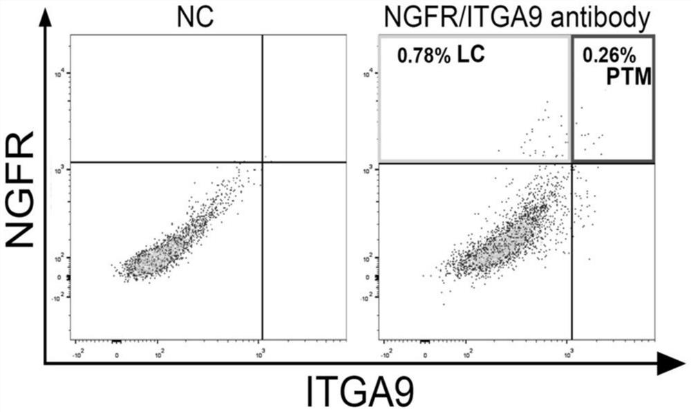
[0072] The present embodiment provides a method for isolating and purifying testis peritubular myoid cells and Leydig cells, the method comprising the following steps:

[0073] (1) Obtain testicular tissue, rinse it with PBS, digest it with collagenase type IV for 15 minutes, use a 100 μm tissue strainer to remove undigested testicular tissue, and keep the cell suspension;

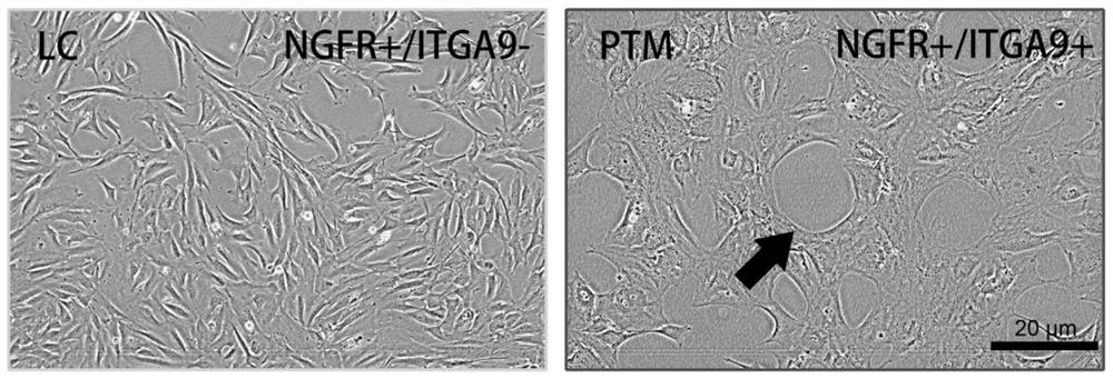
[0074] (2) After centrifuging at 300rpm for 3min and washing the cell suspension twice with PBS, incubate for 10min with the blocking solution in the kit in Example 1, and then use the anti-NGFR protein antibody and anti-ITGA9 protein in the kit in Example 1 at 4°C The antibody was incubated for 30 minutes, washed with PBS and then incubated with the corresponding secondary antibody for 30 minutes at room temperature, and flow cytometry was performed. NGFR positive and ITGA9 negative were Leydig cells (LC), while NGFR and ITGA9 double positive cells were peritubercular myoid cells (PTM) to obtain high-puri...


PUM


Abstract

The invention discloses a biomarker of perimuscular cells of testicular tubes and application of the biomarker. The biomarker comprises an ITGA9 protein. On the basis of the ITGA9 protein, the kit for identifying the testicular perimuscular cells is prepared, the anti-ITGA9 protein antibody is used for specifically recognizing and combining the perimuscular cells, then the testicular perimuscular cells can be effectively identified through methods such as flow analysis and the like, in addition, after the testicular perimuscular cells are identified, the testicular perimuscular cells can be distinguished from Leydig cells, and therefore the identification accuracy of the testicular perimuscular cells is improved. Therefore, the two cells can be effectively separated, the problem that the two cells are difficult to separate by conventional stepwise digestion, differential adherence, gradient centrifugation and other methods is solved, and high-purity Leydig cells and high-purity peritubular muscle-like cells can be obtained through separation and purification.

Description

technical field [0001] The invention belongs to the field of biotechnology, and relates to a biomarker of testicular peritube myoid cells and an application thereof. Background technique [0002] The testis is one of the most complex organs in the human body, including: ① seminiferous tubules composed of peritubular cells, supporting cells, and germ cells at all levels; ② interstitial cells composed of Leydig cells and immune cells; ③ Vasculature composed of endothelial cells and vascular smooth muscle cells, etc. Among them, Leydig cells are the most important cells that produce androgen in men. Dysfunction of Leydig cells can lead to systemic symptoms caused by spermatogenic disorders and androgen decline. A full understanding of the physiological and pathological processes of Leydig cells is of great significance for the study of men's health. In addition, the peritubular myoid cells are also very important for the function of the testis, and they are the main cell compo...

Claims


Application Information

Patent Timeline
IPC IPC(8): C07K14/705G01N33/68G01N33/58C12Q1/02C12N5/071
Inventor 赵亮宇汤育新戴英波叶昆韩厦陈玉琢
Owner THE FIFTH AFFILIATED HOSPITAL SUN YAT SEN UNIV
Why Eureka
Try Eureka