
Method for segmenting a medical data set

a medical data and segmentation technology, applied in image analysis, image enhancement, instruments, etc., can solve the problem of complex manual setting of markings, and achieve the effect of simple method of segmentation

Active Publication Date: 2006-03-16
SIEMENS HEALTHCARE GMBH
View PDF7 Cites 3 Cited by
  • Summary
  • Abstract
  • Description
  • Claims
  • Application Information

AI Technical Summary

Benefits of technology

[0005] An object of the present invention is to provide a simple method for segmentation of data sets.
[0006] This object is achieved in accordance with the invention by a method wherein, in an overview data set, at least one marking is generated with which a data set is measured. The markings limit a relevant region within the overview data set. The segmentation of the measured data set is implemented using the generated markings. The evaluation of the data set (generally composed of many partial images), which is necessary for resetting the markings, is not necessary due to the reuse of the markings. The segmentation thus can be implemented simply and quickly.

Problems solved by technology

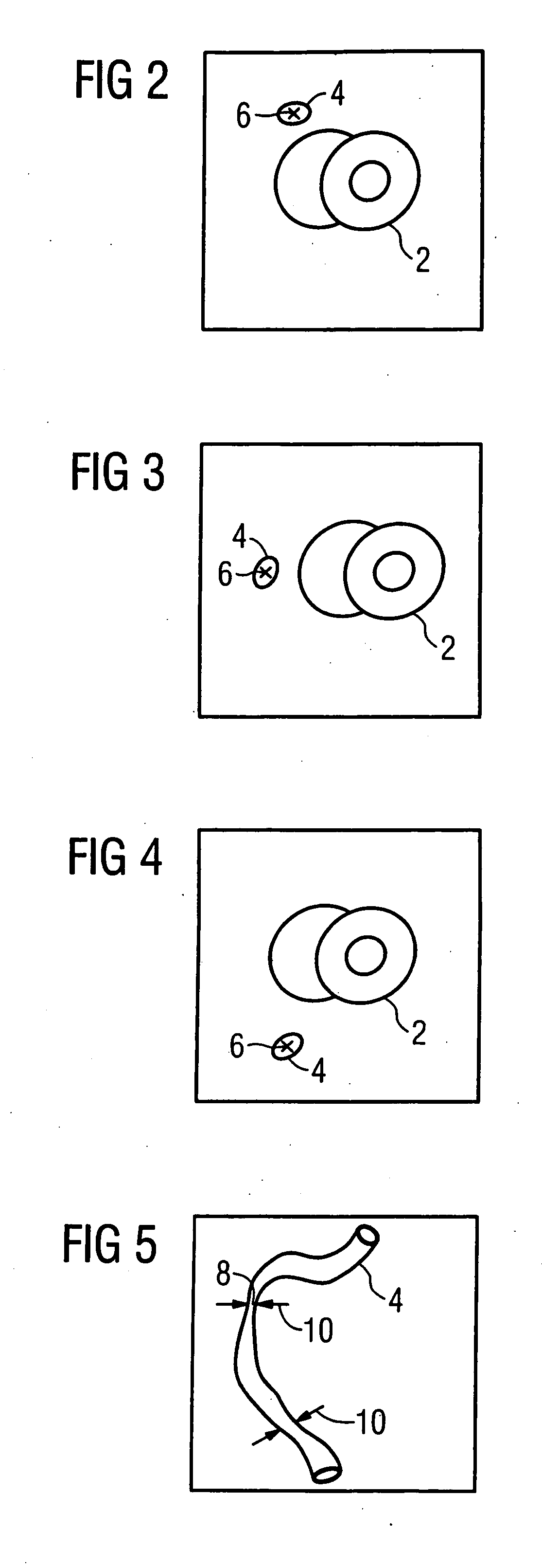
Here a manual setting of markings is complex due to the generally complicated course of the coronary artery in three dimensions.

Method used


Image

  • Method for segmenting a medical data set
  • Method for segmenting a medical data set
  • Method for segmenting a medical data set

Examples


Embodiment Construction

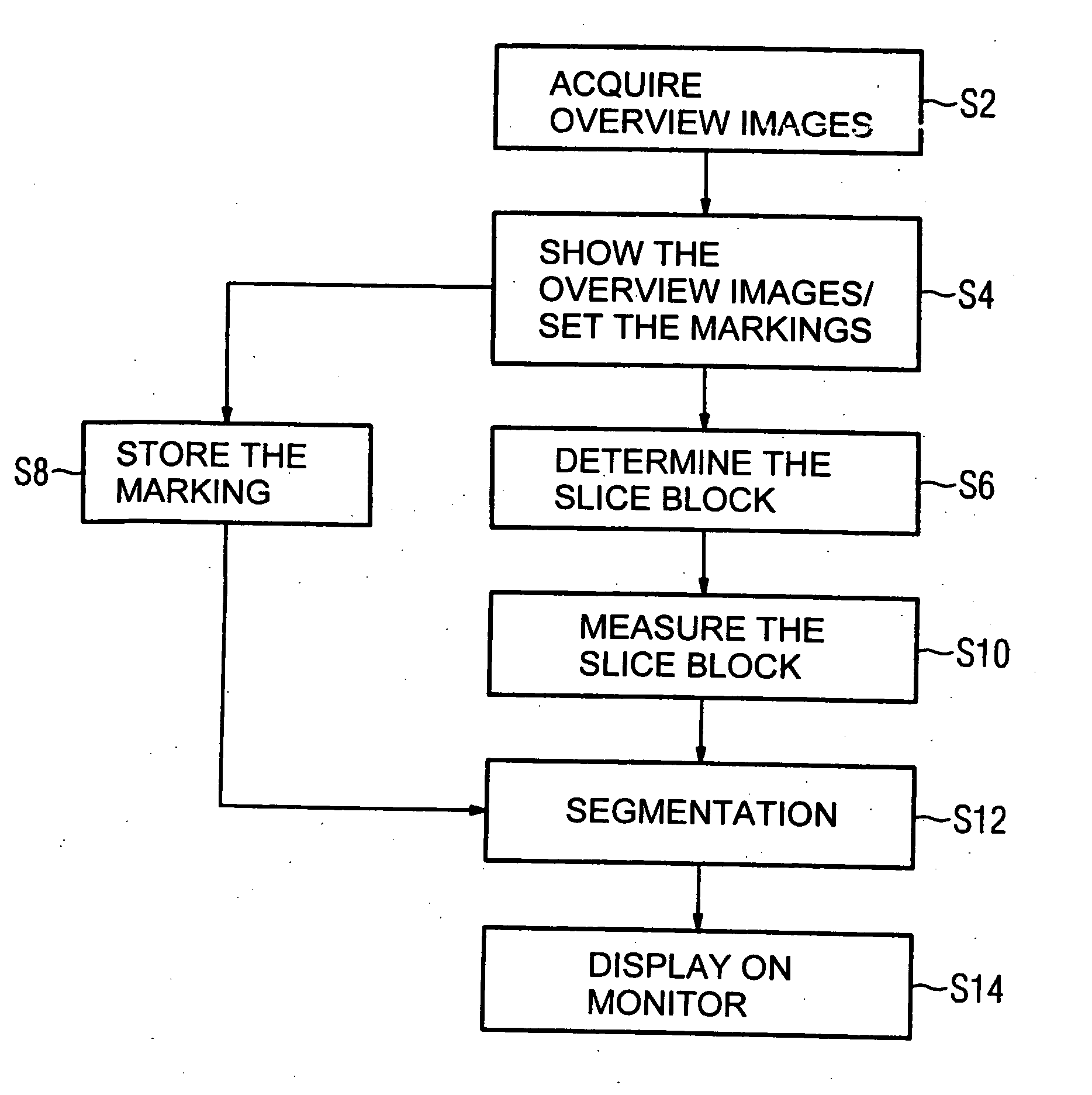
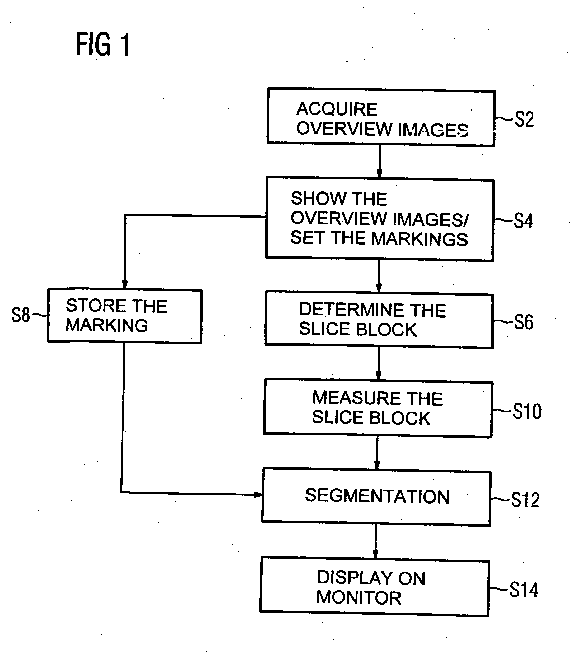
[0011] To examine a coronary artery by magnetic resonance, it is initially necessary to position and orient a slice block to be measured. This slice block is selected so that the coronary artery to be examined, or the part of the coronary artery to be examined, lies within the slice block. For this purpose, in a first method step S2 three overview images in which the coronary artery is to be presented are acquired at various positions. In a second method step S4, each image is shown on a monitor. During the display, a marking is manually generated in each image by an examining physician via an input unit, for example a computer mouse. In a third method step S6, a slice block is determined that optimally maps the region limited by the markings. The markings are stored for further use in a method step S8 parallel to this. The measurement of the determined slice block ensues in a fourth method step S10. The segmentation of the coronary artery to be examined ensues in a fifth method ste...


PUM


Abstract

In a method for segmentation of a medical data set, the segmentation is implemented using markings, the markings being set in the generation of the data set.

Description

BACKGROUND OF THE INVENTION [0001] 1. Field of the Invention [0002] The present invention concerns a method for segmenting a medical data set. [0003] 2. Description of the Prior Art [0004] Segmenting of data sets is necessary in many situations for making a diagnostic finding from medical data sets. A relevant region of a data set is thereby particularly emphasized, or non-relevant regions of a data set are removed. A medical finding is thereby made easier for a physician. To make a finding in the context of a magnetic resonance examination, the physician, for example, can limit the relevant region within a data set by setting a number of markings. The segmentation is implemented using these markings. Thus, for example, the segmenting of a coronary artery proceeds where the markings are placed along the coronary artery and thus mark its course within a three-dimensional data set. After the segmentation, the coronary artery can be disposed, for example, in a pseudo-3D representation,...

Claims


Application Information

Patent Timeline
Patent Type & Authority Applications(United States)
IPC IPC(8): G06K9/34
CPCG01R33/5608G06T7/0081G06T2207/30101G06T2207/20096G06T2207/10088G06T7/11
Inventor ASSMANN, STEFAN
Owner SIEMENS HEALTHCARE GMBH
Learn More