Method for enhancing blood and lymph flow in the extremities
a technology of applied in the field can solve the problems of reducing wound healing ability, neuropathies, poor wound healing, etc., and achieve the effects of enhancing blood and lymph flow
- Summary
- Abstract
- Description
- Claims
- Application Information
AI Technical Summary
Benefits of technology
Problems solved by technology
Method used
Image
Examples
example 1
Patient and Control Subject Screening
[0042]Screening was conducted of consecutive female patients aged 45-70 years who were enrolled in a general internal medicine practice. Patients were excluded with a current fracture of the lower appendicular or axial skeleton, or history of back pain which could be exacerbated by the vibration protocol (see below), known peripheral vascular disease, peripheral neuropathy, uncontrolled hypertension (blood pressure exceeding 150 mm Hg or diastolic blood pressure 95 mm Hg despite treatment), congestive heart failure, diabetes, liver or kidney failure, hyperparathyroidism, multiple myeloma, metastatic carcinoma, Cushing's syndrome, collagen vascular disease, chronic angioedema or lymphedema, uncontrolled hyperthyroidism, chronic substance abuse, or any condition precluding the subject following the protocol or providing informed consent. Subjects with excessive alcohol use (>2 drinks / day) or who smoked were also excluded.
example 2
Laboratory Evaluation
[0043]All experiments started at 9 AM after a brief fast (2 hours). The right arm and right calf blood pressure were monitored intermittently by oscillometry. A vibrating plate (see below) was placed on the footboard of an electrically driven tilt table (Cardiosystems 600, Dallas, Tex., USA). Patients wore rubber soled shoes to ensure electrical isolation and were asked to lie supine with their feet flush with the plate which initially was not oscillating. This situation was designated “0 Hz”. Patients were instrumented to measure blood flow by two forms of measurement: mercury in silastic strain gauge plethysmography (“SGP”) with venous occlusion congestion cuffs and impedance plethysmography (“IPG”). Occlusion cuffs were placed around the lower limb 10 cm above a strain gauge of appropriate size attached to a Whitney-type SGP. Ag / AgCl EKG electrodes for IPG were attached to the left foot and left hand which served as current injectors, and in pairs representin...
example 3
Peripheral Vascular Evaluation by Strain Gauge Plethysmography (“SGP”)
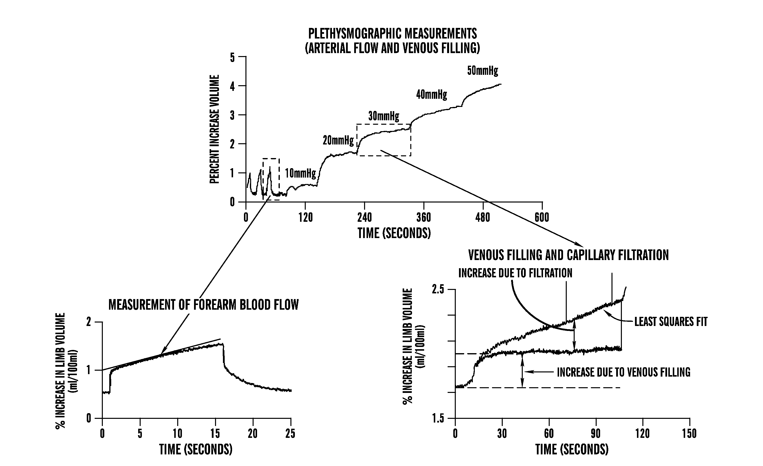
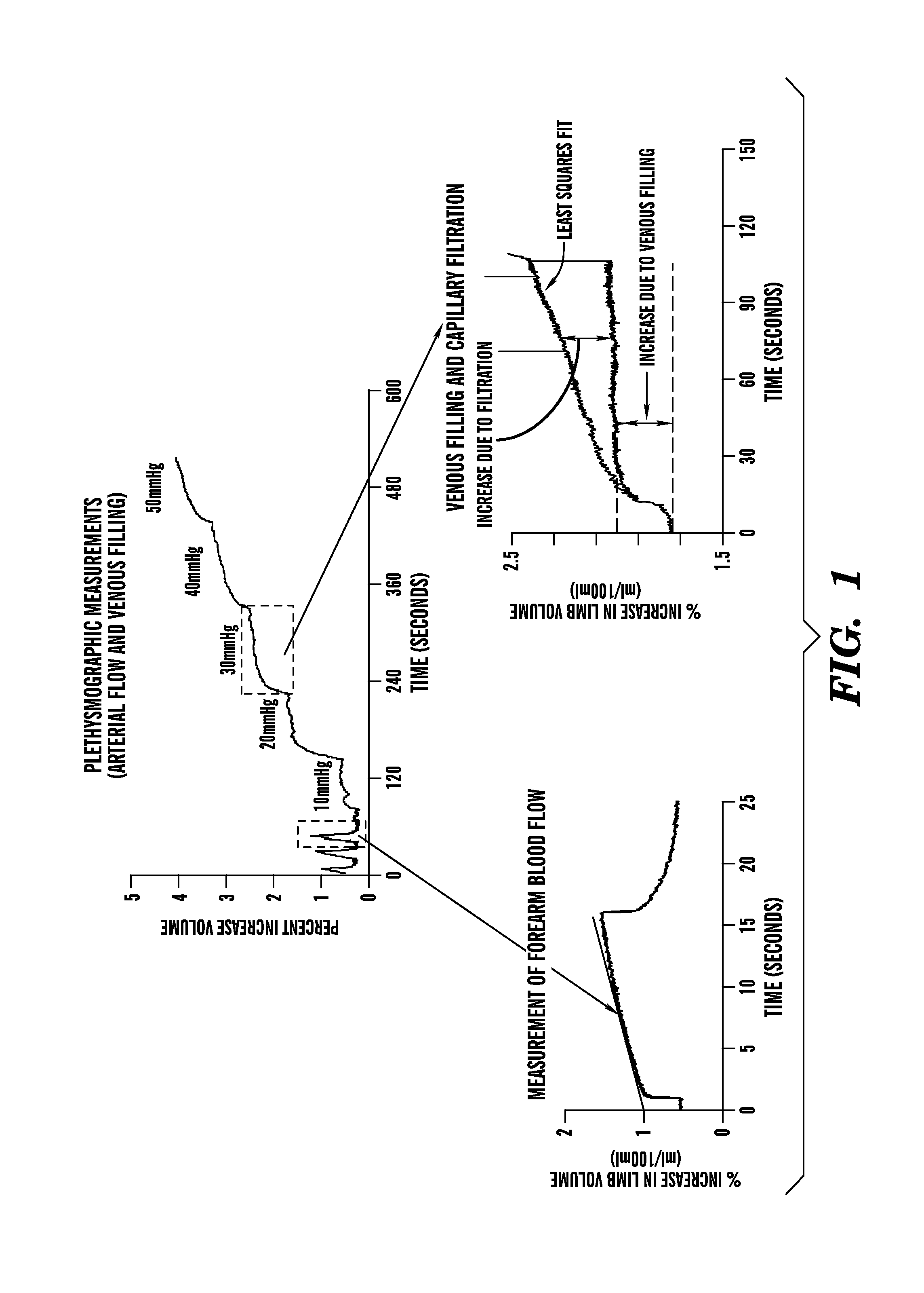
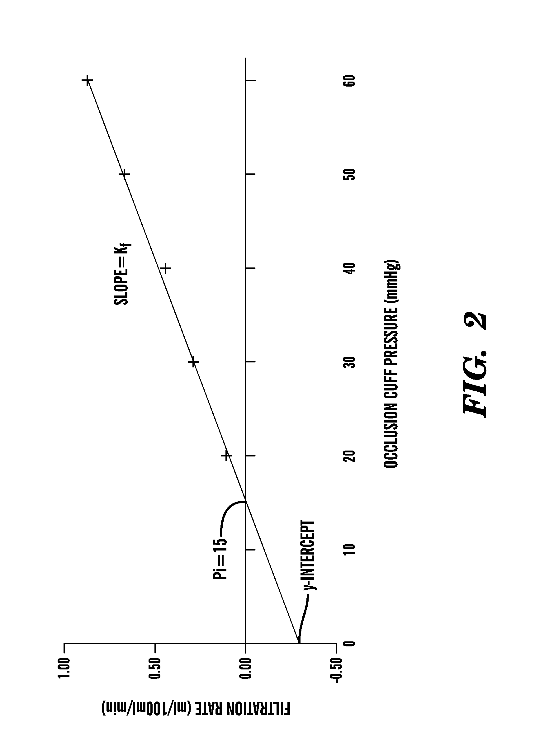
[0045]SGP was used to measure calf blood flows, the calf capacitance vessel pressure (venous pressure, denoted Pv), the calf venous volume-pressure capacitance relation, calf venous capacity and the microvascular filtration (flow-pressure) relation in the supine steady state and during steady state upright tilt to 35° in all subjects. Methods were adapted from the work of Gamble et al. (Gamble et al., “Mercury in Silastic Strain Gauge Plethysmography for the Clinical Assessment of the Microcirculation,”Postgrad Med. J., 68 Suppl. 2:S25-S33 (1992); Gamble et al., “The Effect of Passive Tilting on Microvascular Parameters in the Human Calf: A Strain Gauge Plethysmography Study,”J. Physiol. (London) 498 (Pt. 2):541-552 (1997); Gamble et al., “A Reassessment of Mercury in Silastic Strain Gauge Plethysmography for Microvascular Permeability Assessment in Man,”J. Physiol. (London) 464:407-422 (1993), which are hereby in...
PUM
 Abstract
Description
Claims
Application Information
 - R&D
- Intellectual Property
- Life Sciences
- Materials
- Tech Scout
- Unparalleled Data Quality
- Higher Quality Content
- 60% Fewer Hallucinations
Browse by: Latest US Patents, China's latest patents, Technical Efficacy Thesaurus, Application Domain, Technology Topic, Popular Technical Reports.
© 2025 PatSnap. All rights reserved.Legal|Privacy policy|Modern Slavery Act Transparency Statement|Sitemap|About US| Contact US: help@patsnap.com


