Method for enhancing blood and lymph flow in the extremities

a technology which is applied in the field can solve the problems of neuropathies, and reducing wound healing ability, and achieves the effects of enhancing blood and lymph flow

Inactive Publication Date: 2006-05-25
THE RES FOUND OF STATE UNIV OF NEW YORK
View PDF14 Cites 36 Cited by
  • Summary
  • Abstract
  • Description
  • Claims
  • Application Information

AI Technical Summary

Benefits of technology

[0010] One aspect of the present invention relates to a method of enhancing blood and lymph flow in the lower body of a human subject, which involves applying a stimulus to a plantar surface in the lower body of the subject. The stimulus is effective to displace the skin of the plantar surface between about 10 and 100 microns in amplitude, thereby enhancing the blood and lymph flow in the lower body associated with the stimulated plantar surface.
[0011] Another aspect of the present invention relates to a method of enhancing blood and lymph flow in the upper extremeties of a human subject, which involves applying a stimulus to a palmer surface of the subject. The stimulus is effective to displace the skin of the palmer surface between about 10 and 100 microns in amplitude, thereby enhancing the blood and lymph flow in the extremities associated with the stimulated palmer surface.
[0012] A further aspect of the present invention relates to a method of enhancing blood and lymph flow in the extremities of a human subject, which involves applying a stimulus to one or more plantar or palmer surfaces in an extremity of the subject. The stimulus is effective to displace the skin of the plantar or palmer surface between about 10 and 100 microns in amplitude, thereby enhancing the blood and lymph flow in the extremity associated with the stimulated plantar or palmer surface.
[0013] Yet another aspect of the present invention relates to a method of enhancing blood and lymph flow in the extremities of a human subject, which involves applying an electrical stimulus to one or more plantar or palmer surfaces in an extremity of the subject. The stimulus is effective to directly stimulate cutaneous receptors in the plantar or palmer surface, thereby enhancing blood and lymph flow in the extremity associated with the stimulated cutaneous receptors.
[0014] According to the present invention, plantar and palmer stimulation using electrical, or low level oscillatory, or relatively high frequency vibrational stimulation, requires no connection to the body. Simple contact of the plantar or palmer surface with the stimuli surface is sufficient to trigger somata-sensory detection, with subsequent coupling to the musculature through reflect arcs and central nervous system pathways. Blood and fluid return from the limbs is therefore accomplished through normal skeletal pumping mechanisms. As the required stimulus to trigger this response is minimal, long term use can be prescribed without concern of complications. Moreover, as no “set-up” per se is required for use of the invention, compliance with treatment is much more readily accepted by the user.
[0015] Considering applications to deep vein thrombosis (DVT) alone, the present invention can result in a significant health care costs savings. Orthopaedic surgery, and in particular joint replacement surgery, and the chemotherapy techniques associated with treatment of cancer represent two medical situations where incidence of deep vein thrombosis is remarkably common, though all forms of extended surgery contribute to the incidence rate. 1-2% of patients developing a deep vein thrombosis die from a pulmonary embolism resulting from a DVT, while the remaining patients will incur costs of close to $10,000 per incidence. Over 1,000,000 individuals a year are hospitalized for DVT in the U.S. at a cost exceeding $10 B per year. The present invention represents a relatively low cost approach to significantly reducing or eliminating many of these complications.

Problems solved by technology

Poor blood flow and fluid flow in the extremities results in numerous serious clinical conditions.
In diabetics, reduced blood flow (“arterial insufficiency”) results in ulcerations, reduced wound healing ability, and neuropathies, commonly leading to amputation.
Venous insufficiency (poor venous blood return) leads to edema and poor wound healing.
Lymphatic insufficiency (poor lymphatic return) commonly leads to infection and associated complications.
Pooling of blood in the limbs during upright posture leads to a condition referred to as orthostatic intolerance (“OI”), resulting in a depressed blood pressure and increased heart rate or tachycardia.
Perhaps the most serious complication of reduced blood flow in the extremities, however, is stasis in the lower limbs, and in particular, the deep femoral veins, which can lead to the condition of deep vein thrombosis (“DVT”).
Emboli from such thrombi can break away and lodge in the lungs, often leading to death.
An additional complication of blood and lymph stasis is a reduced ability of tissues to adapt and heal.
Blood stasis can lead to ulceration of tissue and / or failure of skin ulcers to heal, leading to significant complications.
Pressure sores and ulcers are a common complications associated with extended immobility, including sitting and bedrest.
In addition, poor venous and lymphatic return result in the buildup of fluid pressures in the limbs, depriving the bone tissue of necessary nutrient flow, resulting in bone loss (osteoporosis), and as well exacerbates the complications associated with impaired blood flow common in diabetes.
Thus, prevention of blood and lymph stasis is recognized as a critical problem in medicine, home healthcare, long distance travel, and increasingly, even in the workplace.
Convenient means to enhance blood and lymph flow are not currently available.
Current international standards, however, preclude exposing humans to accelerations of this level for more than a few seconds on a daily basis.
In addition, it has been reported, that the application of continuous whole body vibration can, in fact be detrimental to lower body blood flow.
Thus, not only are existing methods and devices uncomfortable, but their chronic use can be irritating to the skin and underlying tissue, and even organ systems, thus decreasing patient compliance and precluding long term use.
In addition, continuous use of prior art methods and apparatus that rely on whole body vibration can, in fact, have a deleterious effect on lower body circulation.

Method used


Image

  • Method for enhancing blood and lymph flow in the extremities
  • Method for enhancing blood and lymph flow in the extremities
  • Method for enhancing blood and lymph flow in the extremities

Examples


example 1

Patient and Control Subject Screening

[0040] Screening was conducted of consecutive female patients aged 45-70 years who were enrolled in a general internal medicine practice. Patients were excluded with a current fracture of the lower appendicular or axial skeleton, or history of back pain which could be exacerbated by the vibration protocol (see below), known peripheral vascular disease, peripheral neuropathy, uncontrolled hypertension (blood pressure exceeding 150 mm Hg or diastolic blood pressure 95 mm Hg despite treatment), congestive heart failure, diabetes, liver or kidney failure, hyperparathyroidism, multiple myeloma, metastatic carcinoma, Cushing's syndrome, collagen vascular disease, chronic angioedema or lymphedema, uncontrolled hyperthyroidism, chronic substance abuse, or any condition precluding the subject following the protocol or providing informed consent. Subjects with excessive alcohol use (>2 drinks / day) or who smoked were also excluded.

example 2

Laboratory Evaluation

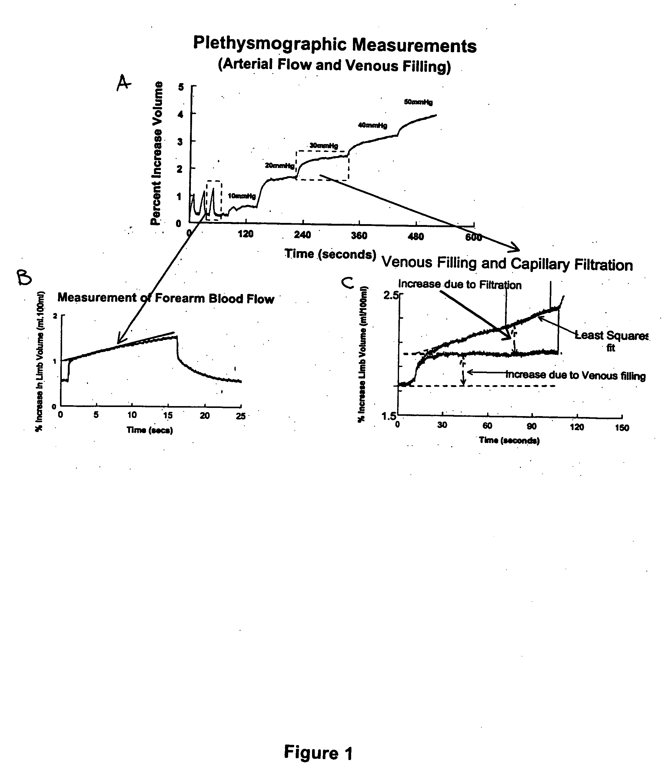
[0041] All experiments started at 9 AM after a brief fast (2 hours). The right arm and right calf blood pressure were monitored intermittently by oscillometry. A vibrating plate (see below) was placed on the footboard of an electrically driven tilt table (Cardiosystems 600, Dallas, Tex., USA). Patients wore rubber soled shoes to ensure electrical isolation and were asked to lie supine with their feet flush with the plate which initially was not oscillating. This situation was designated “0 Hz”. Patients were instrumented to measure blood flow by two forms of measurement: mercury in silastic strain gauge plethysmography (“SGP”) with venous occlusion congestion cuffs and impedance plethysmography (“IPG”). Occlusion cuffs were placed around the lower limb 10 cm above a strain gauge of appropriate size attached to a Whitney-type SGP. Ag / AgCl EKG electrodes for IPG were attached to the left foot and left hand which served as current injectors, and in pairs represent...

example 3

Peripheral Vascular Evaluation by Strain Gauge Plethysmography (“SGP”).

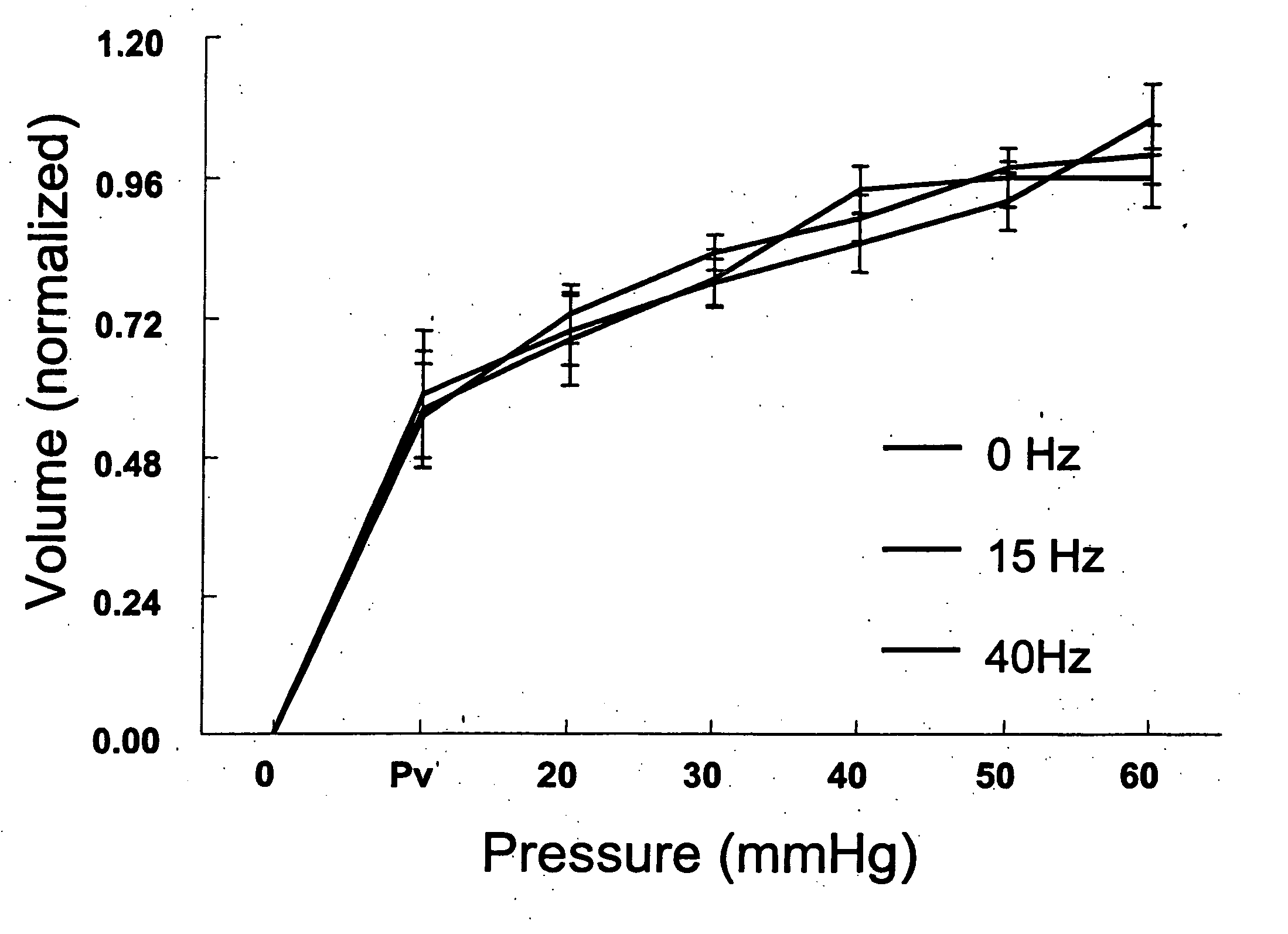
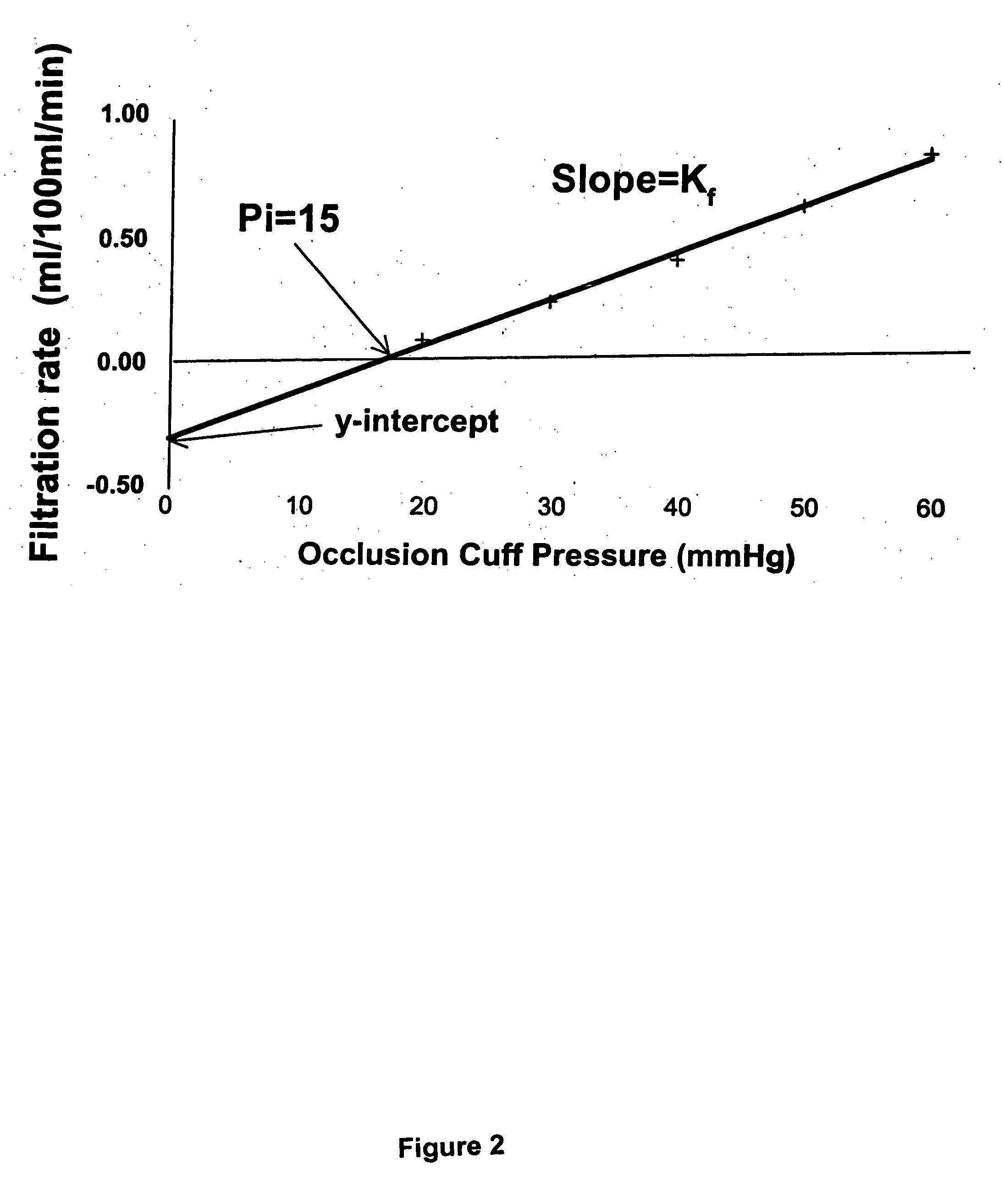
[0043] SGP was used to measure calf blood flows, the calf capacitance vessel pressure (venous pressure, denoted Pv), the calf venous volume-pressure capacitance relation, calf venous capacity and the microvascular filtration (flow-pressure) relation in the supine steady state and during steady state upright tilt to 35° in all subjects. Methods were adapted from the work of Gamble et al. (Gamble et al., “Mercury in Silastic Strain Gauge Plethysmography for the Clinical Assessment of the Microcirculation,”Postgrad Med. J., 68 Suppl. 2:S25-S33 (1992); Gamble et al., “The Effect of Passive Tilting on Microvascular Parameters in the Human Calf: A Strain Gauge Plethysmography Study,”J. Physiol. (London) 498 (Pt. 2):541-552 (1997); Gamble et al., “A Reassessment of Mercury in Silastic Strain Gauge Plethysmography for Microvascular Permeability Assessment in Man,”J. Physiol. (London) 464:407-422 (1993), which are hereby...


PUM


Abstract

Methods for enhancing blood and lymph flow in the extremities of a human subject are disclosed. In one aspect, the methods rely on a stimulus effective to displace the skin of a plantar or palmer surface of the subject, thereby enhancing blood and lymph flow in the extremity associated with the stimulated plantar or palmer surface. In another aspect, the methods rely on an electrical stimulus to directly stimulate cutaneous receptors in a plantar or palmer surface of the subject, thereby enhancing blood and lymph flow in the extremity associated with the stimulated cutaneous receptors. Apparatus for enhancing blood and lymph flow in the extremities of a human subject according to the methods of the present invention are also disclosed.

Description

[0001] The subject matter of this application was made with support from the United States Government under National Heart Lung and Blood Institute of the National Institutes of Health, Grant No. 1RO1HL66007. The U.S. Government may have certain rights.FIELD OF THE INVENTION [0002] The present invention relates to methods for enhancing blood and lymph flow in the lower body and upper extremeties of a human subject. BACKGROUND OF THE INVENTION [0003] Poor blood flow and fluid flow in the extremities results in numerous serious clinical conditions. In diabetics, reduced blood flow (“arterial insufficiency”) results in ulcerations, reduced wound healing ability, and neuropathies, commonly leading to amputation. Venous insufficiency (poor venous blood return) leads to edema and poor wound healing. Lymphatic insufficiency (poor lymphatic return) commonly leads to infection and associated complications. Pooling of blood in the limbs during upright posture leads to a condition referred to ...

Claims


Application Information

Patent Timeline
Patent Type & Authority Applications(United States)
IPC IPC(8): A61H1/00
CPCA61H23/02A61H2023/0209A61H2205/12A61N1/36003A61N1/36021A61H2209/00
Inventor MCLEOD, KENNETH J.
Owner THE RES FOUND OF STATE UNIV OF NEW YORK
Why Eureka
Try Eureka