Raman spectral analysis for disease detection and monitoring

a raman spectral analysis and disease detection technology, applied in the field of raman spectroscopy, can solve the problems of complex diagnostic procedures, high cost, and high labor intensity of current diagnostic procedures for cancer and other diseases

Inactive Publication Date: 2012-12-13
VUKELIC SINISA
View PDF2 Cites 10 Cited by
  • Summary
  • Abstract
  • Description
  • Claims
  • Application Information

AI Technical Summary

Benefits of technology

[0024]FIG. 10e is a graphical illustration of analysis of a selected segment of Raman spectra of a low-grade prostate cancer tissue sample
[0025]FIG. 10f is a graphical illustration of analysis of a selected segment of Raman spectra of a high-grade prostate cancer tissue sample
[0026]The novel aspects of the invention as to organization and method of use, together with further objects and advantages thereof, will be better understood from the following disclosure considered in connection with the accompanying drawings in which one or more preferred embodiments of the invention are illustrated by way of example. It is to be expressly understood, however, that the detailed description and drawings are for the purpose of illustration and description only and are not intended as a definition of the limits of the invention.
[0027]As used herein, the term “comprises” refers to a part or parts of a whole, but does not exclude other parts. That is, the term “comprises” is open language that requires the presence of the recited element or structure or its equivalent, but does not exclude the presence of other elements or structures. The term “comprises” has the same meaning and is interchangeable with the terms “includes” and “has”. The term “set” has the meaning of one or more of said elements. The term “tissue” is intended to include both human and animal tissue, including individual cells, whether in situ or removed as a sample. Furthermore, any use of the term “or” as used herein is generally intended to mean “and / or” unless otherwise indicated.

Problems solved by technology

Current diagnostic procedures for cancer and other diseases are complex, often cumbersome and usually require highly trained personnel.
Because careful tissue preparation is required, this process is relatively slow and arduous.
Such subtle differences can be difficult or time-consuming to detect, even for highly experienced pathologists.

Method used


Image

  • Raman spectral analysis for disease detection and monitoring
  • Raman spectral analysis for disease detection and monitoring
  • Raman spectral analysis for disease detection and monitoring

Examples


example

[0079]The following example is provided for the purposes of illustration only and the disclosure is not limited to the specific method followed in the example.

[0080]Histopathological slides containing biopsied prostate tissue from human subjects were screened using Raman spectroscopy. Raman spectra were obtained from biopsied specimens of healthy individuals and individuals diagnosed with prostate adenocarcinoma. The samples were acquired from the Cooperative Human Tissue Network (CHTN) of the National Institutes of Health (NIH). The measurements were obtained using an assembly comprising a confocal Raman spectrometer consisting of Research Electro Optics He-NE laser emitting 632.8 nm wavelength with 30 mW maximum output power, a Nikon Eclipse TE300 inverted optical microscope, a Horiba i320 triple grating spectrometer and a deep cooled Synapse TE detector.

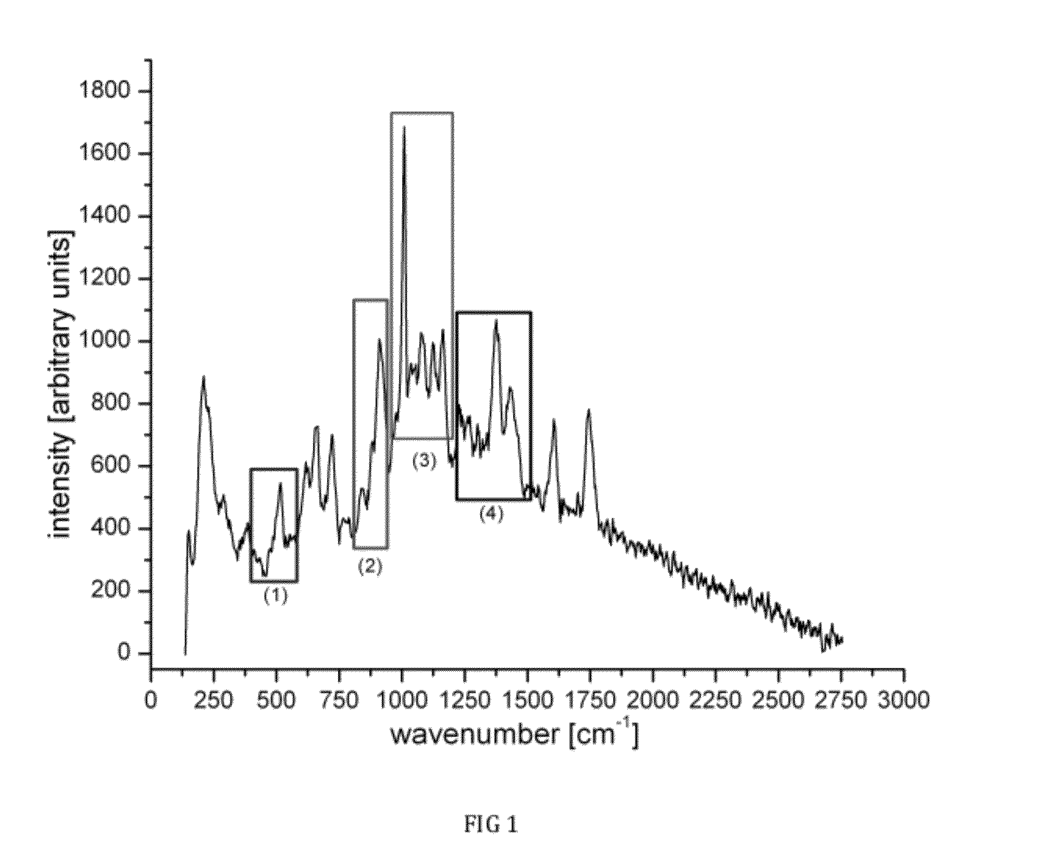
[0081]FIG. 9 shows superimposed Raman spectra of normal tissue, low grade prostate cancer and high grade prostate adenocarcinoma...


PUM


Abstract

A system and method for Raman spectral analysis for diagnostic purposes to identify the disease state of biological tissue. The sampled tissue can be any human or animal tissue, including but not limited to prostate, breast, cervical tissue, etc. Raman spectroscopy is used to provide Raman bands which are decomposed and their chemical fingerprints identified after mathematical modeling. The method enables identification of chemical compounds in the tissue being diagnosed. Comparison with reference samples permits inferences concerning the presence or absence of disease, the stage of disease, and/or the response of disease to treatment. The method provides sufficient information about the chemical composition of the tissue to differentiate between normal and abnormal states, and may also reveal the locations of abnormalities. The method may also be used to monitor the treatment of a subject and/or the response of disease to drugs or other treatments.

Description

FIELD OF INVENTION[0001]The present disclosure relates generally to methods of Raman spectroscopy, and, in particular Raman spectral analysis for diagnostic purposes to detect and monitor potential or actual disease state of human or animal tissue.BACKGROUND OF THE INVENTION[0002]Raman spectroscopy provides information about the vibrational state of molecules. Many molecules have atomic bonds capable of existing in a number of vibrational states. Such molecules are able to absorb incident radiation that matches a transition between two of the allowed vibrational states and to subsequently emit radiation. Most often, absorbed radiation is re-radiated at the same wavelength, a process designated Rayleigh or elastic scattering. In some instances, the re-radiated radiation can contain slightly more or slightly less energy than the absorbed radiation (depending on the allowed vibrational states and the initial and final vibrational states of the molecule). The energy difference between t...

Claims


Application Information

Patent Timeline
Patent Type & Authority Applications(United States)
IPC IPC(8): A61B6/00G01J3/44
CPCG01N21/65
Inventor VUKELIC, SINISA
Owner VUKELIC SINISA
Why Eureka
Try Eureka