Method and apparatus for metal artifact reduction in 3D X-ray image reconstruction using artifact spatial information

a technology of artifacts and spatial information, applied in the field of image reconstruction, can solve the problems of streak artifacts in the formation of such fluoroscopic images, and achieve the effect of reducing or minimizing metal-related streak artifacts

Active Publication Date: 2008-05-06
STRYKER EURO OPERATIONS HLDG LLC
View PDF8 Cites 26 Cited by
  • Summary
  • Abstract
  • Description
  • Claims
  • Application Information

AI Technical Summary

Benefits of technology

[0007]One embodiment of the present invention relates to an image reconstruction device, system or machine. More particularly, one embodiment relates to a 3D X-ray image reconstruction method and apparatus that reduces or minimizes metal-related streak artifacts.

Problems solved by technology

However, it is known that metal implants such as pedicle screws and fracture plates / pins for example, may generate streak artifacts in the formation of such fluoroscopic images.
Such streak artifacts are created due to the sharp difference in signal attenuation at the boundary of the metal implants and the patient's anatomy.
Such ramp filters may mitigate but do not eliminate the streak artifacts, since the filters are generally broadly applied to the entire scan data set.

Method used


Image

  • Method and apparatus for metal artifact reduction in 3D X-ray image reconstruction using artifact spatial information
  • Method and apparatus for metal artifact reduction in 3D X-ray image reconstruction using artifact spatial information
  • Method and apparatus for metal artifact reduction in 3D X-ray image reconstruction using artifact spatial information

Examples


Embodiment Construction

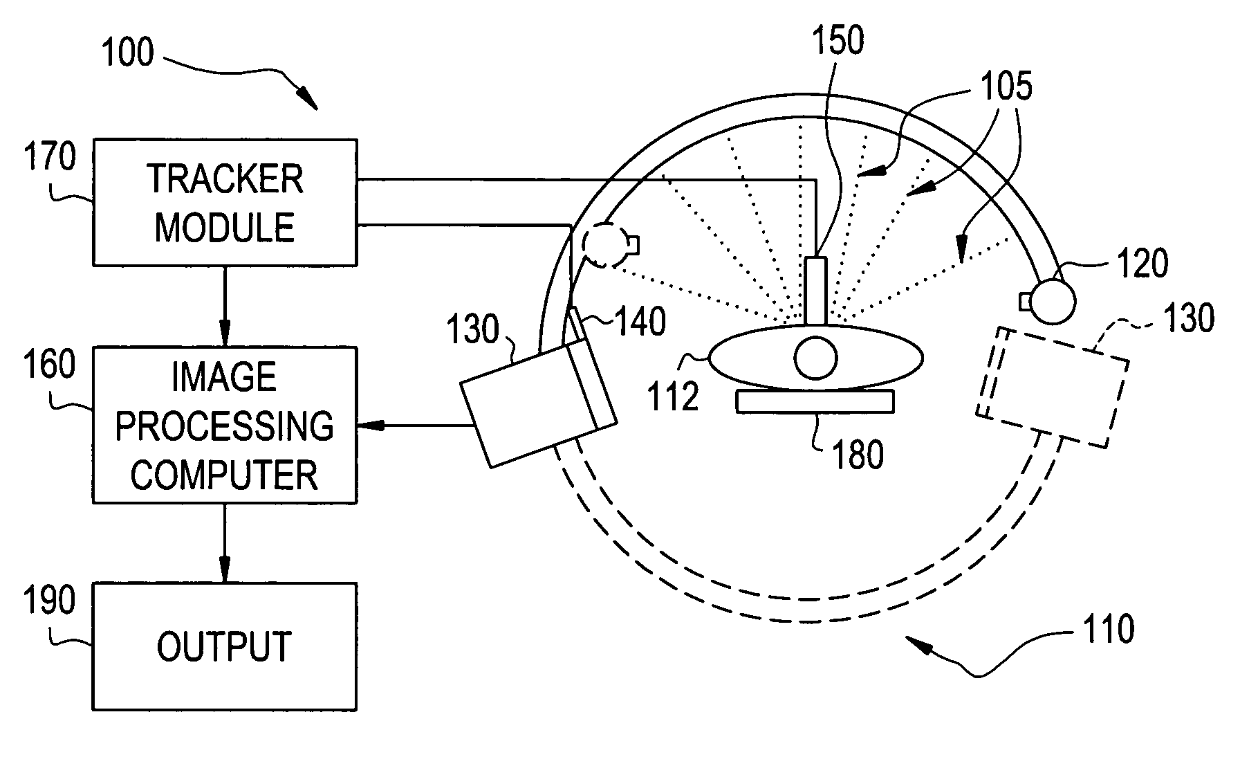
[0020]For the purpose of illustration only, the following detailed description references a certain embodiments of an X-ray or fluoroscopic image apparatus, system, device or machine. However, it is understood that the present invention may be used with other devices or imaging systems.

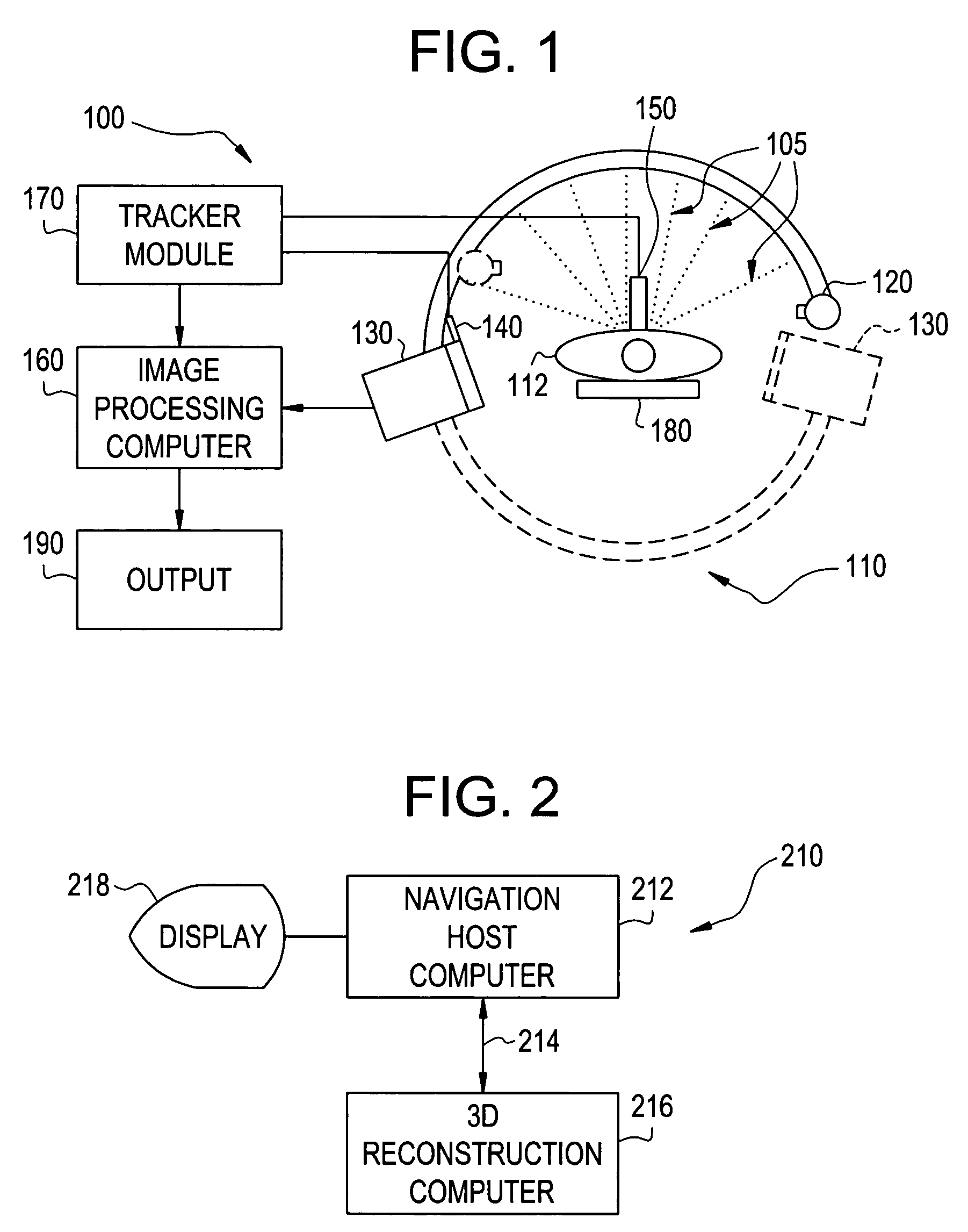
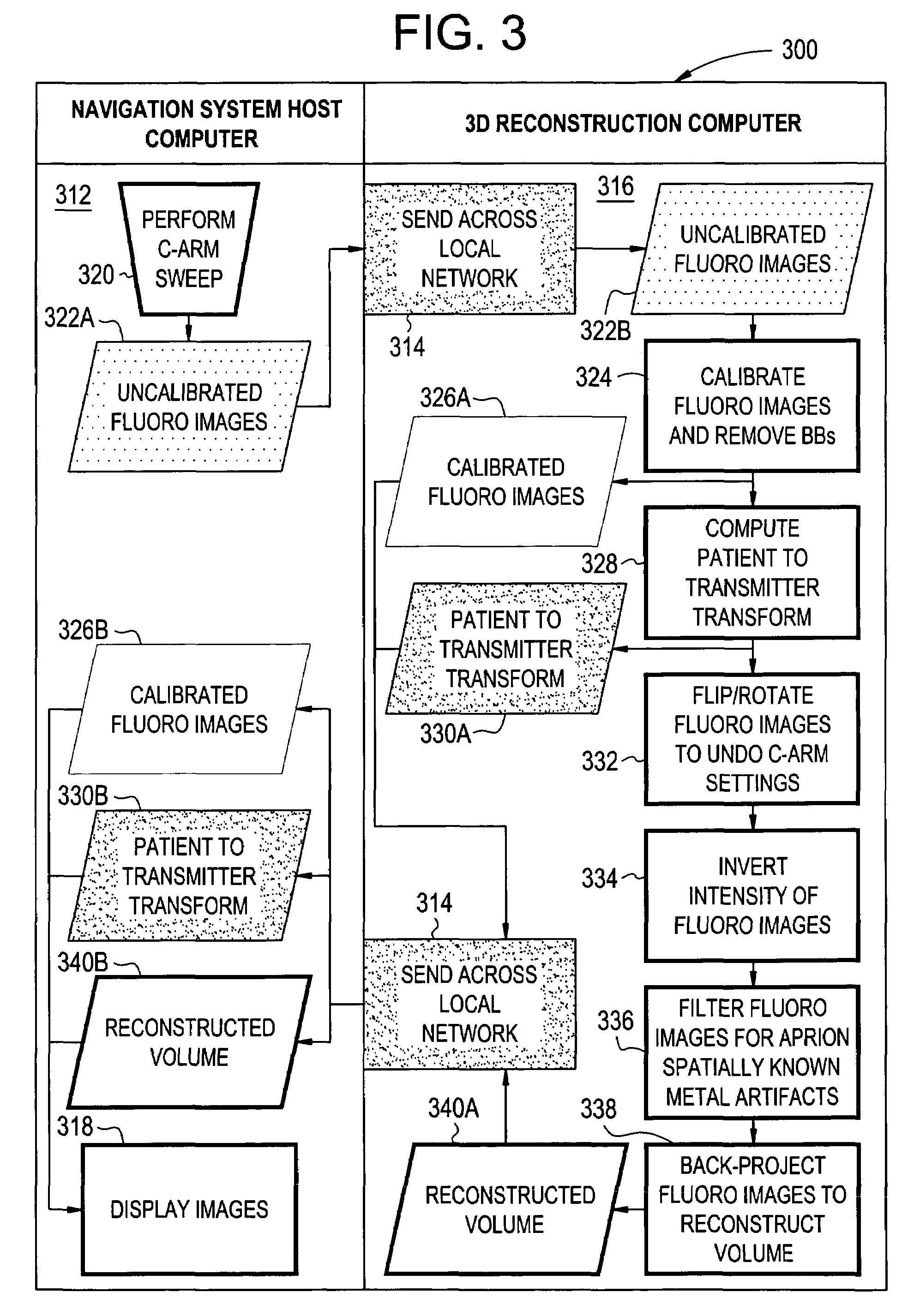
[0021]3D fluoroscopic images may be generated from 2D fluoroscopic projection images by the image device or system illustrated in FIGS. 1, 2 and 3, using cone beam reconstruction techniques similar to those discussed below. The inherent position and / or orientation information for each 2D projection taken or acquired by the C-arm helps ensure the spatial relationship between the plurality of 2D projection images used to create 3D images. Since the tracking information is, in at least one embodiment, dynamically referenced to the 2D images, embodiments are enabled to directly locate one or more metal implants in each of the 2D images. In one embodiment, a pointer may be used to identify the location of ...


PUM


Abstract

The present invention relates to a method and apparatus for reducing metal artifact in image reconstruction. In at least one embodiment, the method for reducing metal artifacts in image reconstruction comprises collecting at least one uncalibrated image (an X-ray or fluoroscopic image for example) and calibrating the at least one uncalibrated image, forming at least one calibrated fluoroscopic image. A patient to transmitter transform is computed using at least the at least one calibrated image. A reconstructed volume is formed using at least a patient to transmitter transform. At least two of the at least one calibrated fluoroscopic image, the patient to transmitter transform and the reconstructed volume may be combined, forming at least one display image. This image may then be displayed, using a display device for example.

Description

RELATED APPLICATIONS[0001][Not Applicable]FEDERALLY SPONSORED RESEARCH OR DEVELOPMENT[0002][Not Applicable]MICROFICEH / COPYRIGHT REFERENCE[0003][Not Applicable]BACKGROUND OF THE INVENTION[0004]This application is directed in general to image reconstruction. More particularly, this application is directed to metal artifact reduction in an image reconstruction using artifact spatial information.[0005]Forming CT and 3D Fluoroscopic images of a patient's anatomy is well known in the art. It should be appreciated that it is desirable for surgeons to see how well internal fixation of metal implants was performed in post surgical images. However, it is known that metal implants such as pedicle screws and fracture plates / pins for example, may generate streak artifacts in the formation of such fluoroscopic images. Such streak artifacts are created due to the sharp difference in signal attenuation at the boundary of the metal implants and the patient's anatomy.[0006]Many techniques or methods ...

Claims


Application Information

Patent Timeline
Patent Type & Authority Patents(United States)
IPC IPC(8): G06K9/00
CPCA61B6/032A61B6/5252G06T5/002G06T2200/04G06T2207/10081G06T2207/10121A61B6/5258A61B6/00
Inventor ZETTEL, HUBERT A.GROSZMANN, DANIEL E.KAPUR, TINA
Owner STRYKER EURO OPERATIONS HLDG LLC
Why Eureka
Try Eureka