
MART-1 T cell receptors
a t cell receptor and mart cell technology, applied in the field of mart cell receptors, can solve problems such as difficult implementation of procedures
- Summary
- Abstract
- Description
- Claims
- Application Information
AI Technical Summary
Benefits of technology
Problems solved by technology
Method used
Image

Examples
example 1
Cloning of the MART-1 T-Cell Receptor (TCR)
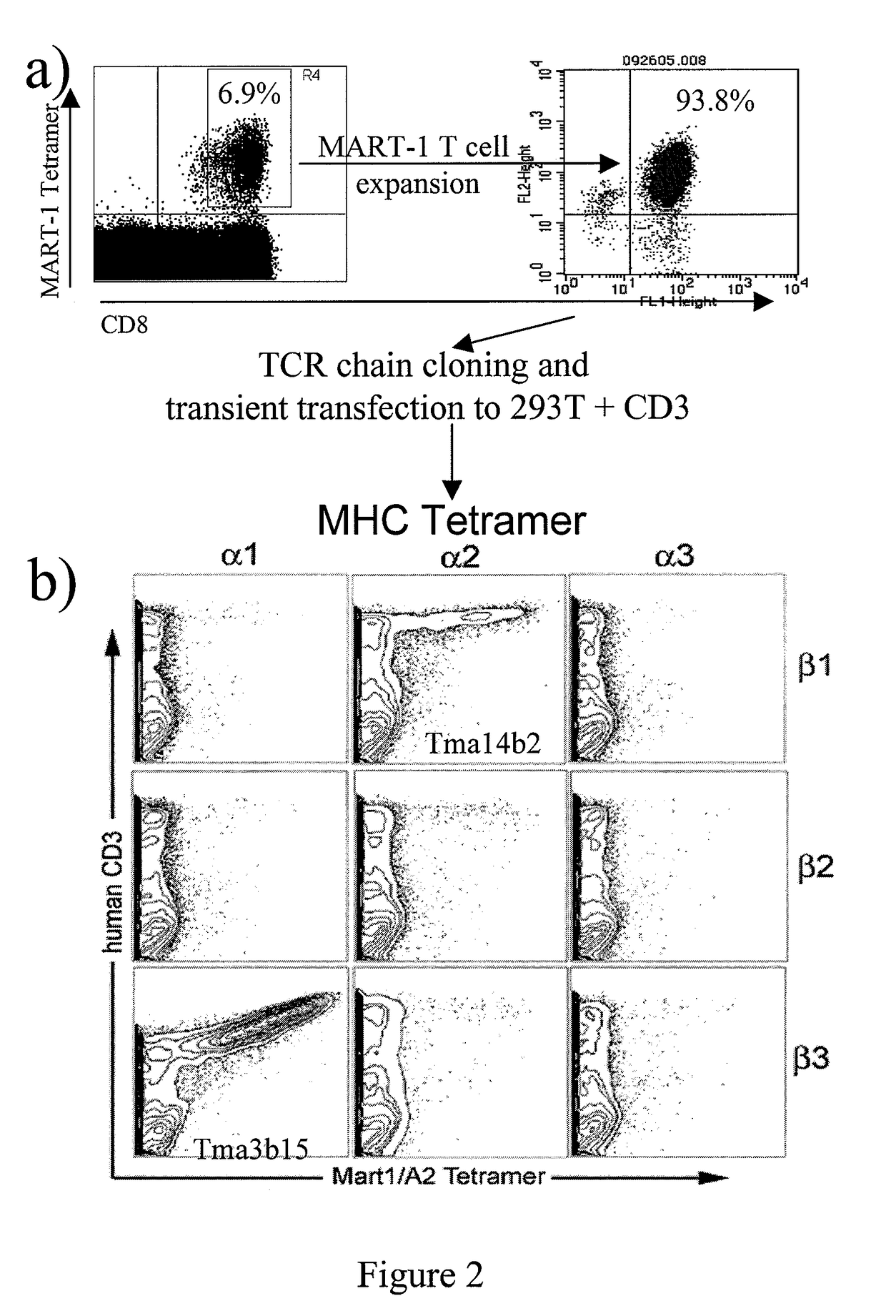
[0240]The MART-1 TCR gene was cloned from a patient with an unusual population of high affinity MART-1-specific T cells representing 5-7% of the patient's total CD8+ cells (FIG. 2). The particular patient is a 7-year survivor of widely metastatic melanoma to lung, nodes and brain who was treated with MART-1 antigen-transduced dendritic cells (DCs) and CTLA-4-blocking monoclonal antibody (CP-675,206). The patient had an unusually favorable clinical course, presenting with no evidence of disease (NED) 7 years after lung, brain and nodal metastases from melanoma.
[0241]Purified CD8+ T cells from the patient were stimulated by DCs pulsed with the MART-126-35 peptide (SEQ ID NO: 20) and expanded in IL-15. The cells were tetramer-sorted, and total RNA was isolated. 5-prime Race RT-PCR was employed to amplify the variable region of the TCR. Several TCR alpha chains and beta chains were identified. Genes encoding these chains were then cloned into m...
example 2
Construction of a MART-1 TCR Lentiviral Vector
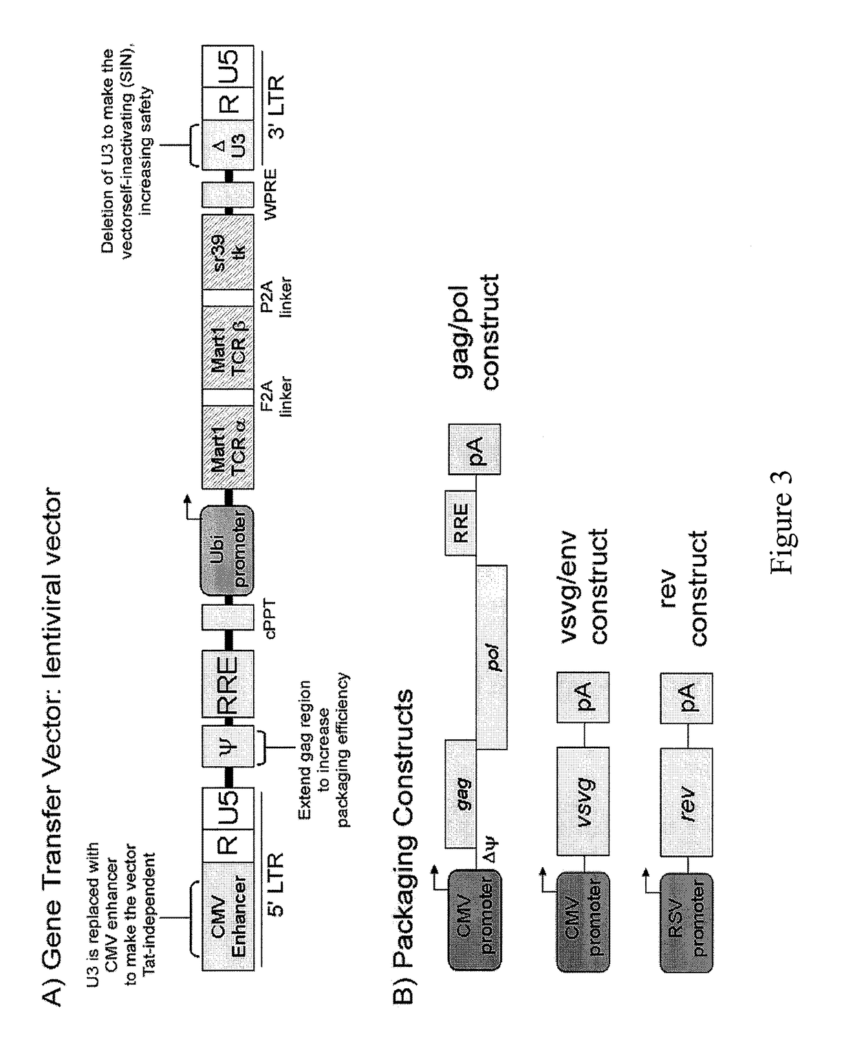
[0243]The gene transfer vector used in this study is a third generation HIV-based lentiviral vector (FIG. 3). FIG. 3 illustrates the MART-1 lentiviral vector used in experimental studies, known henceforth as “FFUW-M1-TCR / sr39tk.” (SEQ ID NO: 17).
[0244]Extensive modifications were introduced into the vector to improve its safety for use. For example, most of the U3 region of the 3′ LTR has been deleted to ensure that transcription from the 5′ LTR is efficiently suppressed after reverse transcription and integration into cellular genome. As a result, the integrated vector cannot generate full-length vector RNA (called self-inactivating or SIN). In addition, an enhancer region derived from cytomegalovirus (CMV) immediate early promoter was used to replace the U3 region of 5′ LTR. This modification eliminates the requirement for the presence of Tat protein for vector production without compromising viral titer. Furthermore, a human ubiquitin...
example 3
Functional Activity of TCR-Engineered Cells
[0247]FUW-M1-TCR / sr39tk lentivirus vector transduction results in the surface expression of a MART-1-specific TCR in modified Jurkat cells and PBMC. In this experiment, Jurkat cells were transduced with the lentiviral vector encoding MART-1 TCR with a F2A linker (vector FUW-M1-TCR / sr39tk); resulting cells were designated as Jurkat-M1. Tetramer staining confirmed that over 70% cells are MART-1 positive (FIG. 5a). The assay for measuring interleukin-2 (IL-2) release was performed by co-incubation of Jurkat-M1 with T2 cells pulsed with various concentration of MART-126-35 peptide (designated as T2 / M1) for three days. Supernatants were then collected for IL-2 ELISA analysis. Upon stimulation with MART-126-35 peptide, the Jurkat-M1 cells responded efficiently as manifested by healthy IL-2 production; the IL-2 response was correlated with the dosage of peptide supplied to the cells. Lack of IL-2 production from various controls demonstrated the s...
PUM
| Property | Measurement | Unit |
|---|---|---|
| concentration | aaaaa | aaaaa |
| size | aaaaa | aaaaa |
| affinity | aaaaa | aaaaa |
Abstract
Description
Claims
Application Information