Method for inducing and differentiating amniotic epithelial stem cells into functional liver cells and application of method

A technology for inducing differentiation and stem cells, applied in cell dissociation methods, cell culture active agents, biochemical equipment and methods, etc., can solve the problems of stem cell tumorigenicity, limited sources of bone marrow and umbilical cord blood, and difficult clinical use of cells, etc. Achieve the effects of inhibiting transaminase activity, restoring liver albumin levels, and enriching cell sources

Active Publication Date: 2018-12-25
NANCHANG UNIV
View PDF5 Cites 5 Cited by
  • Summary
  • Abstract
  • Description
  • Claims
  • Application Information

AI Technical Summary

Problems solved by technology

However, the limited sources of bone marrow and umbilical cord blood, the ethical issues and tumorigenicity of embryonic stem ce

Method used


Image

  • Method for inducing and differentiating amniotic epithelial stem cells into functional liver cells and application of method
  • Method for inducing and differentiating amniotic epithelial stem cells into functional liver cells and application of method
  • Method for inducing and differentiating amniotic epithelial stem cells into functional liver cells and application of method

Examples


Embodiment 1

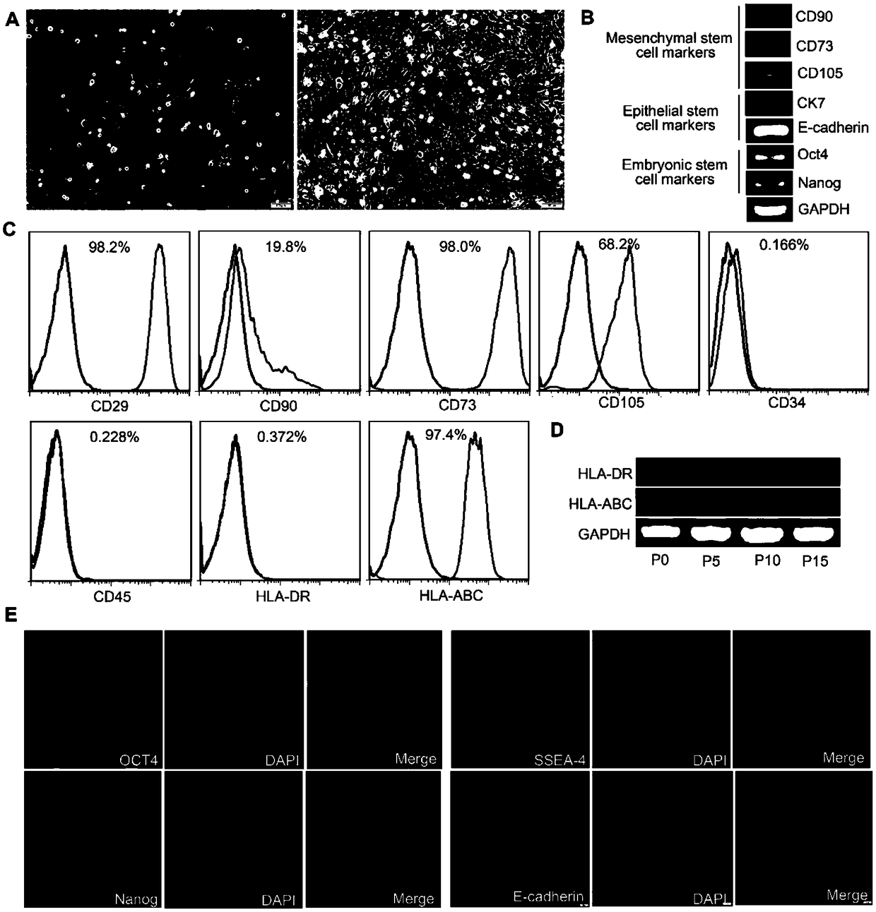
[0089] Example 1. Isolation, culture, expansion and GFP marker of amniotic membrane epithelial stem cells (HAESCs):

[0090] Under the premise of obtaining the consent of the newborn's family members, the fresh amniotic membrane was isolated. After 2 hours of antibiotic treatment, microbiological detection and safety detection of infectious disease pathogens were carried out. After the amniotic membrane was digested by trypsin / EDTA (0.25%) in a water bath at 37°C for 1 hour, stop solution was added. After centrifugation, the supernatant was removed, and the amnion epithelial stem cell culture medium was placed in 5% CO 2 , 95% humidity, and cultured in a 37°C incubator. Change the medium after 2-3 days to remove unattached cells, and then change the medium every 2 days. When the confluence of the cells reached 80%, trypsin / EDTA was used for passage. The amniotic epithelial stem cells of passage 3 were transfected with GFP using lentivirus. Immunofluorescence and flow cyto...

Embodiment 2

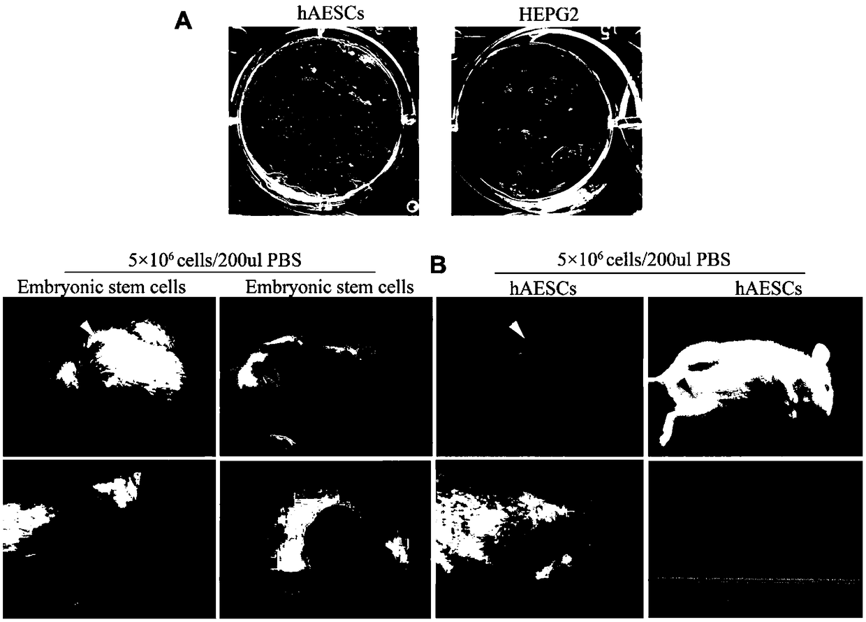
[0176] Example 2. In vivo and in vitro tumorigenicity detection of amniotic membrane epithelial stem cells

[0177] 1. In vivo and in vitro tumorigenicity detection of amniotic membrane epithelial stem cells

[0178]1) Take the amniotic membrane epithelial cells with good growth condition in the third generation of culture, showing vigorous growth, large cell bodies, clear nuclei, abundant cytoplasm, and strong refraction under the microscope. ) digestion, when the cells turned into a single circle under the microscope, the digestion was terminated with DMEM medium containing 10% (volume concentration) fetal bovine serum, gently pipetted and centrifuged to obtain cell pellets, washed with PBS (PBS without calcium and magnesium ions). (the purpose is to wash off trypsin, fetal bovine serum and other substances);

[0179] 2) the amniotic membrane epithelial stem cells in step 1) were inoculated on soft agar, and after culturing for 30 days, the colony formation was observed;

...

Embodiment 3

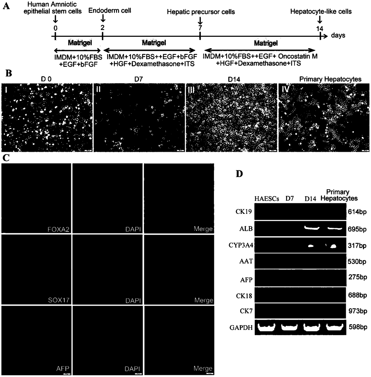
[0187] Example 3. Induction and differentiation of amniotic epithelial stem cells into hepatocytes and their functional detection

[0188] The specific operation procedures are as follows:

[0189] 1. In vitro directional induction of amniotic epithelial stem cells to differentiate into hepatocytes

[0190] 1) Take the well-grown cells of the third generation of culture and digest them with trypsin-EDTA digestion solution (0.25%). DMEM medium to terminate the digestion, gently pipetting and centrifugation to obtain cell pellets, washed twice with PBS (PBS washing solution without calcium and magnesium ions) (the purpose is to wash away substances such as trypsin and fetal bovine serum);

[0191] 2) The above-mentioned cell pellets washed with PBS were inoculated into a 6-well plate pretreated with Matrigel for induction culture, and the inoculated cell density was 3 × 10 5 1 / well, add 2ml of amniotic epithelial stem cell universal medium to each well, at 37°C, 5% CO 2 , cul...


PUM


Abstract

The invention discloses a method for inducing and differentiating human amniotic epithelial stem cells into liver cells with biological functions. The method includes the steps: 1) separating, culturing, amplifying and identifying the human amniotic epithelial stem cells, and marking the human amniotic epithelial stem cells by GFP (green fluorescent protein); 2) in-vitro induction differentiation:using third-seventh-generation human amniotic epithelial stem cells with GFP markers for test, selecting a first system containing inducing media I, II and III or a second system containing inducingmedia IV, V and VI to perform induced differentiation, and inducing and differentiating the cells by the first system (inducing time is 14-15 days) or the second system (inducting time is 22-23 days)to obtain liver sample cells which can express specific markers of the liver cells and have normal liver cell functions; 3) in-vivo transplantation: transplanting the liver sample cells generated by inducing into a body of an acute liver injury mouse. Acute liver injury can be obviously relieved, the liver function of the mouse is restored, and the survival rate of the mouse is increased.

Description

technical field [0001] The invention relates to a method for inducing differentiation of liver cells using amniotic membrane epithelial stem cells. Background technique [0002] Orthotopic liver transplantation is the gold standard for the treatment of end-stage liver disease. However, the shortage of liver sources is the biggest bottleneck for clinical hepatocyte transplantation. Therefore, finding a suitable and abundant source of hepatocytes is a hot research topic in the field of biology. In 1981, Evans et al. established the first mouse ES cell line, which was used as a model to confirm that mouse ES cells can not only be induced to differentiate into hepatic progenitor cells in vitro, but also further differentiate into biologically functional hepatocytes. Animal experiments have shown that when the hepatocytes differentiated in vitro are transplanted into liver injury model mice, the transplanted cells can be fused to the liver tissue and relieve the liver injury of...

Claims


Application Information

Patent Timeline
IPC IPC(8): C12N5/071
CPCC12N5/067C12N2500/24C12N2500/32C12N2500/44C12N2501/11C12N2501/115C12N2501/12C12N2501/33C12N2509/00
Inventor 辛洪波柳全文李婧嫄刘倩玉任康康
Owner NANCHANG UNIV
Why Eureka
Try Eureka