A method for quality control of ultrasound section images of fetuses in the second and third trimesters

A section image, fetal technology, applied in the directions of ultrasound/sound/infrasound image/data processing, image enhancement, image analysis, etc., can solve the problems of inconsistent quality control results, large manpower and material resources, difficult to popularize

Active Publication Date: 2021-10-29
深圳蓝湘智影科技有限公司
View PDF15 Cites 0 Cited by
  • Summary
  • Abstract
  • Description
  • Claims
  • Application Information

AI Technical Summary

Problems solved by technology

[0005] In view of the above deficiencies or improvement needs of the prior art, the present invention provides a method for quality control of ultrasound section images of fetuses in the middle and late pregnancy. Material resources lead to technical problems that are difficult to be widely applied in actual clinical practice, and the technical problems of inconsistency in quality control results caused by different doctors' perceptions of whether fetal ultrasound section images are standard or not. The present invention can assist ultrasound doctors Nearly 40 more standard and higher-quality fetal sections were selected (including thalamic horizontal section, lateral ventricle horizontal section, cerebellar horizontal section, nasolabial coronal section, double eyeball horizontal section, facial median sagittal section, Four-chamber heart view, left ventricular outflow tract view, right ventricular outflow tract view, three-vessel view, three-vessel tracheal view, diaphragm coronal view, diaphragm sagittal view, upper abdominal transverse view, umbilical cord abdominal wall entrance abdominal transverse view, umbilical artery level Transverse section of bladder, transverse section of both kidneys, sagittal section of both kidneys, coronal section of both kidneys, sagittal section of spine, transverse section of spine, coronal section of spine, horizontal transverse section of scapula, long axis section of left and right humerus, left and right ulna-radius long-axis section, left and right ulna-radius short-axis section, ilium level transverse section, left and right femur long-axis section, left and right tibia-fibula long-axis section, left and right Right tibia and fibula short-axis section, sagittal coronal section of both hands, sagittal coronal section of both feet, sagittal section of internal os of pregnant women, section of umbilical cord placental entrance, section for measuring placental thickness, umbilical artery blood flow spectrum, and section of the largest amniotic fluid pool )image

Method used


Image

  • A method for quality control of ultrasound section images of fetuses in the second and third trimesters
  • A method for quality control of ultrasound section images of fetuses in the second and third trimesters
  • A method for quality control of ultrasound section images of fetuses in the second and third trimesters

Examples


Embodiment Construction

[0084] In order to make the object, technical solution and advantages of the present invention clearer, the present invention will be further described in detail below in conjunction with the accompanying drawings and embodiments. It should be understood that the specific embodiments described here are only used to explain the present invention, not to limit the present invention. In addition, the technical features involved in the various embodiments of the present invention described below can be combined with each other as long as they do not constitute a conflict with each other.
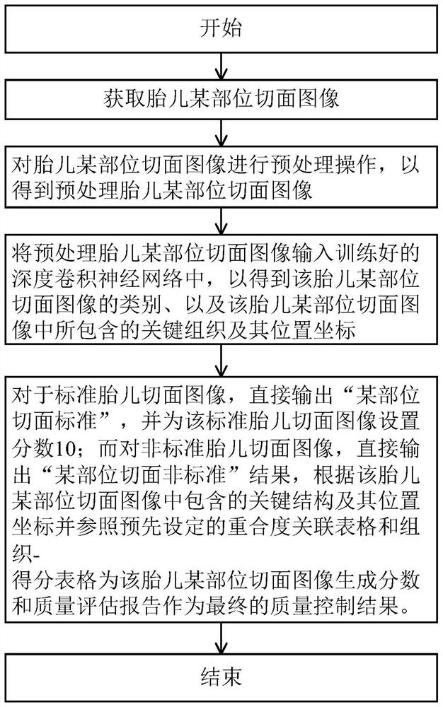
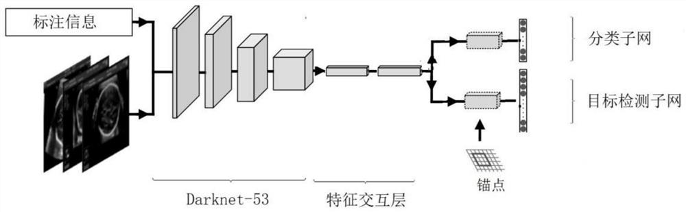
[0085] The basic idea of ​​the present invention is to provide a method for quality control of ultrasound section images of various parts of the fetus in the middle and late pregnancy (nearly 40 parts in total), and use a computer to learn the Combined with the experience and knowledge of ultrasound experts, combined with the quality control standards for section images of various parts of the f...


PUM


Abstract

The invention discloses a method for quality control of an ultrasound slice image of a fetus in the second and third trimesters. , input the preprocessed fetal part section image into the trained deep convolutional neural network to obtain the category of the fetal section section image, as well as the key structure and its position coordinates contained in the fetal section section image, for For the section image of a certain part of the fetus whose category is standard, the result of "standard section of a certain part" is directly output. For the section image of a certain part of the fetus whose category is non-standard, the result of "non-standard section of a certain part" is directly output, and the reason for the non-standard section is pointed out. . A score and a quality assessment report are generated for the sliced ​​images of a certain part of the fetus. The invention can solve the technical problem that the existing method is difficult to widely popularize and apply in actual clinical practice.

Description

technical field [0001] The invention belongs to the technical field of prenatal ultrasonic examination, and more specifically relates to a method for quality control of ultrasonic section images of fetuses in the middle and late pregnancy. Background technique [0002] At present, fetal ultrasonography in the second and third trimesters is the preferred examination method for prenatal diagnosis and screening of defective infants. However, due to the uncertainty of fetal position, maternal body mass index, doctor's operation techniques, ultrasound physical characteristics, fetal movement and other factors, there are often great differences in the clarity and standardization of fetal ultrasound images obtained by ultrasound examination. Greatly affected the accuracy of the test results. Therefore, it is necessary to carry out quality control (Quality Control, QC for short) on fetal ultrasound section images. [0003] The existing method for quality control of fetal ultrasoni...

Claims


Application Information

Patent Timeline
Patent Type & Authority Patents(China)
IPC IPC(8): A61B8/08A61B8/00G06T7/00
CPCA61B8/0866A61B8/5215G06T7/0002G06T7/0012G06T2207/10132G06T2207/20081G06T2207/20084G06T2207/30044G06T2207/30168
Inventor 李肯立李胜利谭光华文华轩
Owner 深圳蓝湘智影科技有限公司
Why Eureka
Try Eureka