Protective device for radioactive source in radiology department, and use method

A protective device and radioactive source technology, applied in the field of radiology, can solve the problems of medical personnel injury, inconvenient automatic deployment, etc., and achieve the effect of convenient automatic protection and avoiding manual pulling

Inactive Publication Date: 2022-01-07
青岛山大齐鲁医院(山东大学齐鲁医院(青岛))
View PDF0 Cites 0 Cited by
  • Summary
  • Abstract
  • Description
  • Claims
  • Application Information

AI Technical Summary

Problems solved by technology

[0005] Aiming at the deficiencies of the prior art, the present invention provides a protective device and a method of use for radioactive sources in the radiology department, which has the advantages of being convenient for automatic deployment and reducing the harm caused by the entry of medical personnel, and solves the problem of some radioactive sources in the radiology department. For radiation, the protective cover is used for protection, and most of them are manually pulled and deployed by medical staff. It is inconvenient to automatically deploy and cause serious injuries to medical staff.

Method used


Image

  • Protective device for radioactive source in radiology department, and use method
  • Protective device for radioactive source in radiology department, and use method
  • Protective device for radioactive source in radiology department, and use method

Examples


Embodiment Construction

[0031] The following will clearly and completely describe the technical solutions in the embodiments of the present invention with reference to the accompanying drawings in the embodiments of the present invention. Obviously, the described embodiments are only some, not all, embodiments of the present invention. Based on the embodiments of the present invention, all other embodiments obtained by persons of ordinary skill in the art without making creative efforts belong to the protection scope of the present invention.

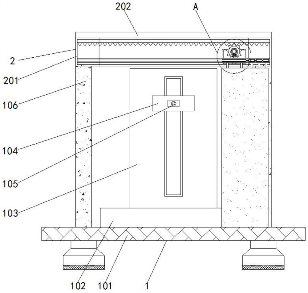
[0032] see Figure 1-2 , the protection device and method of use for radioactive sources in the radiology department in this embodiment include a support mechanism 1, and the top of the support mechanism 1 is fixedly connected with a protection mechanism 2;

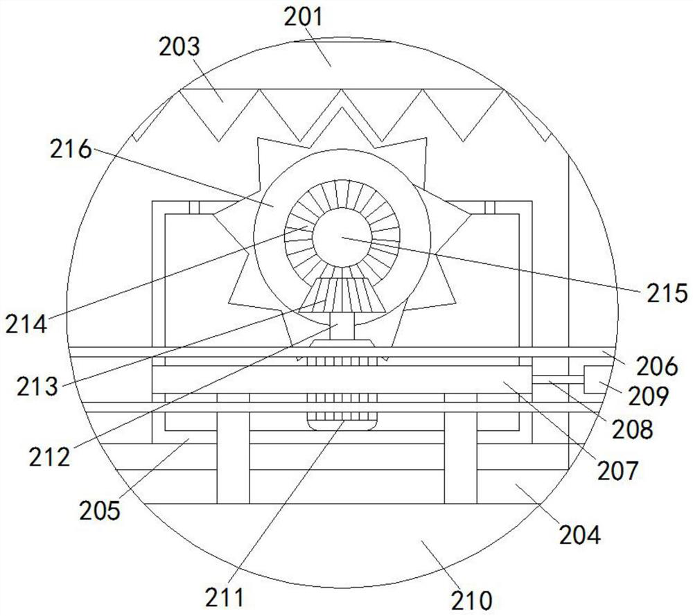
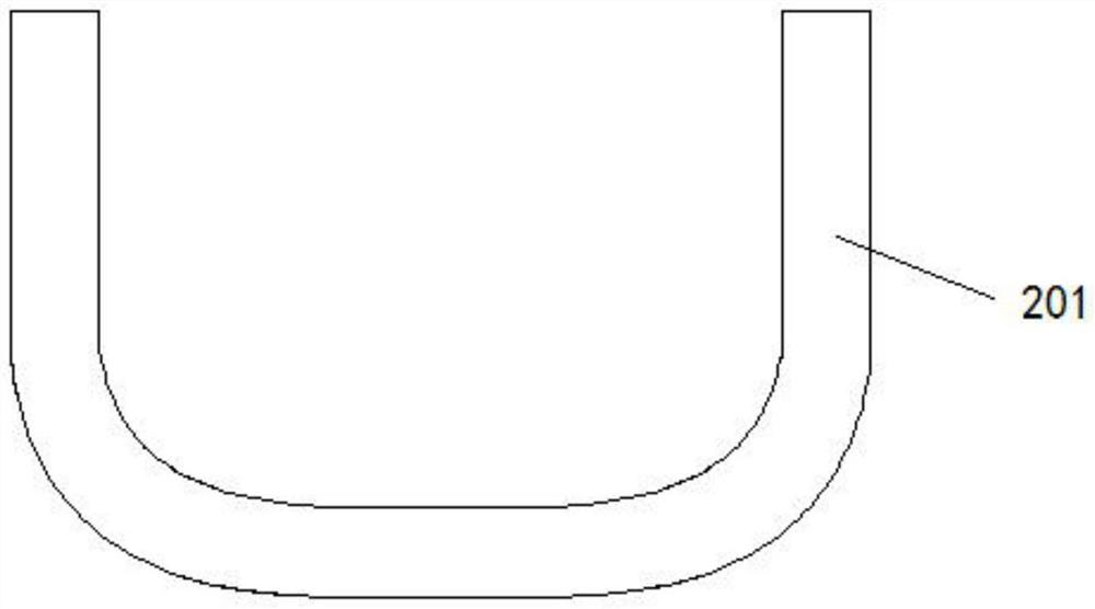
[0033] The protection mechanism 2 comprises a chute body 201, the top of the chute body 201 is fixedly connected with a protective cover 202, the inner wall of the chute body 201 is fixedly connected with...


PUM


Abstract

The invention relates to the technical field of the radiology department, and discloses a protective device for a radioactive source in the radiology department, and a use method. The protective device comprises a supporting mechanism, wherein the top of the supporting mechanism is fixedly connected with a protection mechanism; the protection mechanism comprises a sliding groove body, wherein the top of the sliding groove body is fixedly connected with a protection cover; the inner wall of the sliding groove body is fixedly connected with a rack; the bottom of the sliding groove body is provided with a fixing groove; the inner wall of the sliding groove body is movably connected with a box body; the inner wall of the sliding groove body is fixedly connected with a guide rail; a sliding body is fixedly connected to the outer wall of the box body; a pulling rope is fixedly connected to the right side of the sliding body; a sliding block is fixedly connected to one end, which is far away from the sliding body, of the pulling rope; a protective cover is fixedly connected to the bottom of the sliding body; and a motor is fixedly connected in the box body. The protection device for the radioactive source in the radiology department, and the use method have the advantages that automatic unfolding is facilitated, harm caused by entering of medical staff is reduced and the like.

Description

technical field [0001] The invention relates to the technical field of radiology, in particular to a protective device and a use method for radioactive sources in the radiology department. Background technique [0002] The Department of Radiology is an important auxiliary examination department of the hospital. In the construction of modern hospitals, the Department of Radiology is a department that integrates inspection, diagnosis and treatment. Many diseases in various clinical departments must be diagnosed and assisted by radiology equipment inspection. Radiology equipment generally includes ordinary X-ray film machines, computerized X-ray photography systems, direct digital X-ray photography systems, computerized X-ray tomography, nuclear magnetic resonance, digital subtraction angiography systems, etc. [0003] Some radioactive sources in the radiology department are radiated, and protective covers are used for protection. Most of them are manually pulled and deployed b...

Claims


Application Information

Patent Timeline
Patent Type & Authority Applications(China)
IPC IPC(8): A61B6/10
CPCA61B6/107
Inventor 杨志成冷珊珊
Owner 青岛山大齐鲁医院(山东大学齐鲁医院(青岛))
Why Eureka
Try Eureka