
Method for analyzing retinal fundus images and related products

A fundus image and retina technology, applied in the field of image processing, can solve the problems of single measurement results, discontinuous topology, and inability to perform refined detection of key points.

Active Publication Date: 2022-06-21
BEIJING AIRDOC TECH CO LTD
View PDF10 Cites 0 Cited by
  • Summary
  • Abstract
  • Description
  • Claims
  • Application Information

AI Technical Summary

Problems solved by technology

If the arteries and veins segmented using the traditional segmentation network (for example, the output layer shares parameters and uses a multi-category softmax activation function), there are topological discontinuities, and then the key points cannot be identified based on the discontinuous topological map. Refined detection makes the measurement result single and lacks practicability

Method used


Image

  • Method for analyzing retinal fundus images and related products
  • Method for analyzing retinal fundus images and related products
  • Method for analyzing retinal fundus images and related products

Examples


Embodiment Construction

[0034] The technical solutions in the embodiments of the present invention will be clearly and completely described below with reference to the accompanying drawings in the embodiments of the present invention. Obviously, the described embodiments are part of the embodiments of the present invention, not all of the embodiments. Based on the embodiments of the present invention, all other embodiments obtained by those skilled in the art without creative efforts shall fall within the protection scope of the present invention.

[0035] It should be understood that the terms "first", "second", "third" and "fourth" in the claims, description and drawings of the present invention are used to distinguish different objects, rather than to describe a specific order . The terms "comprising" and "comprising" used in the description and claims of the present invention indicate the presence of the described features, integers, steps, operations, elements and / or components, but do not exclu...


PUM


Abstract

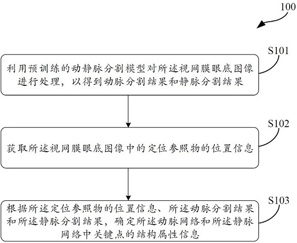
The invention relates to a method for analyzing retinal fundus images and related products. Wherein the method includes: using a pre-trained arteriovenous segmentation model to process the retinal fundus image to obtain an arterial segmentation result and a vein segmentation result, wherein the arterial segmentation result includes a continuous arterial network, and the vein segmentation result includes A continuous vein network; acquiring position information of a positioning reference object in the retinal fundus image; and determining the arterial network and the position information according to the position information of the positioning reference object, the artery segmentation result, and the vein segmentation result The structural attribute information of the key points in the vein network. Through the technical solution of the present invention, using the continuous arterial network and venous network of the retinal fundus image, combined with the position information of the positioning reference object, the refined detection of the multi-dimensional structural attributes of the key points can be realized.

Description

technical field [0001] The present invention generally relates to the technical field of image processing. More particularly, the present invention relates to a method for analyzing retinal fundus images, as well as devices and computer-readable storage media for performing the aforementioned method. Background technique [0002] This section is intended to provide a background or context for the embodiments of the invention that are recited in the claims. The descriptions herein may include concepts that may be explored, but not necessarily concepts that have been previously thought of or explored. Therefore, unless otherwise indicated herein, what is described in this section is not prior art to the description and claims in this application and is not admitted to be prior art by inclusion in this section. [0003] Cardiovascular disease is a disease that seriously threatens human health, and early identification of individuals at high risk of cardiovascular disease is t...

Claims


Application Information

Patent Timeline
Patent Type & Authority Patents(China)
IPC IPC(8): G06T7/00G06T7/11G06N3/04
Inventor 黄烨霖赵昕
Owner BEIJING AIRDOC TECH CO LTD
Learn More