Due to the tortuous anatomy and the three-dimensional arch structures anchoring stents using existing techniques requires a steep learning curve and extensive monitoring may result in adverse radiation exposure for the patient.
Besides embolism, several complications can occur during carotid access, including dissection of the carotid arteries caused by the ledge effect due to unopposed space between a larger guide catheter (7-9 F) and a smaller diagnostic catheter and guide-wire-related branch arterial perforations.
The small branch perforations can be deadly, due to the rapid development of retropharyngeal bleeding and airway compromise particularly with an anti-coagulated patient.
The most dreaded complication associated with the remote guide catheter access method is guide catheter prolapse, with the carotid filter getting entangled in the stent and detaching or embolizing.
Additionally, cerebral embolism during carotid access is a risk during a carotid stent procedure.
Additionally, access to the carotid and vertebral arteries, as well as occasionally to other vessels, can sometimes be impossible to achieve in routine transfemoral fashion in certain anatomical situations.
This is especially difficult with type III aortic arches.
However, in many anatomical situations, access to some vessels can be difficult.
If the catheter does not have adequate physical support to resist such recoil and displacement, it will fall out of the desired position.
In some cases this could prevent appropriate access to the desired pathological region altogether.
In other cases, it can create complications during a procedure when catheters and devices suddenly recoil and displacement and are dislodged from the desired location.
Such movement is undesirable.
However, there is a general reluctance to puncture the right brachial artery due to the need to navigate through the innominate artery and arch and due to the risk for complications such as direct nerve trauma and ischemic occlusion resulting in long-term disability (AlvarezTostado J. A. et al.
Distal thrombectomy is a technically difficult procedure (Singh P. et al.
Despite good clinical outcome, limitations of this device include operator learning curve, the need to traverse the occluded artery to deploy the device distal to the occlusion, the duration required to perform multiple passes with device, clot fragmentation and passage of an embolus within the bloodstream (Meyers P. M. et al.
These stents are not ideal for treating intracranial disease due to their rigidity which makes navigation in the convoluted intracranial vessels difficult (Singh P. et al.
Drawbacks of this method include delayed in-stent thrombosis, the use of platelet inhibitors which may cause intracerebral hemorrhage (ICH) and perforator occlusion from relocation of the thrombus after stent placement (Samaniego E. A. et al Front Neurol.
These include the requirement for double anti-platelet medication, which potentially adds to the risk of hemorrhagic complications and the risk of in-stent thrombosis or stenosis.
Despite the potential to diminish procedure time and to improve recanalization rates, drawbacks to using these devices remain.
Although mechanical endovascular neurointerventions using a transfemoral approach are the current standard for the treatment of acute stroke, it is difficult to access the right internal carotid artery and right vertebral artery via these transfemoral techniques when certain aortic arch variation occurs.
A similar transfemoral access problem can occur when vertebral arteries arise at an acute angle from the subclavian artery, or in some variations of the left internal carotid or other vessels.
Although results have improved, repair of this abnormality is associated with a significant mortality and morbidity (Tchervenkov C. I. et al.
For example, the acute angle at which the left common carotid artery branches from the aortic arch in the bovine arch configuration makes mechanical endovascular neurointervention difficult, especially when additional tortuosity (i.e., twists) in the aorta and / or the carotid artery are present.
However, when a wire is advanced through these catheters in order to achieve distal access to the artery head, these catheters lack adequate support which results in recoil and displacement into the aortic arch of the advancing wire.
The lack of adequate support and the resulting recoil and displacement of the advancing wire make effective treatment impossible.
Even when catheterization is achieved in these situations, the process of arriving at the correct combination of catheters and wires results in long treatment delays.
In cases of acute stroke, long delays in obtaining access to arteries often leads to additional irreversible cell death with additional permanent neurologic injury.
Additionally, with aging the anatomy of the takeoff of the great vessels often changes, which can make access to the cerebral vasculature more difficult.
This is particularly problematic as with aging the incidence of major thromboembolic strokes, for which emergent endovascular thrombectomy is often the best treatment, also increases The branching pattern of the brachiocephalic branch from the aortic arch was categorized into three types based on the vertical distance from the origin of the brachiocephalic branch to the top of the arch, which determined the arch type.
However, the Simmons has two significant short comings: first these catheters only maintain their shape when they are unconstrained and second, passing a wire through a Simmons it will result changing the shape of the Simmons and alter the direction of the hole at the end of the Simmons.

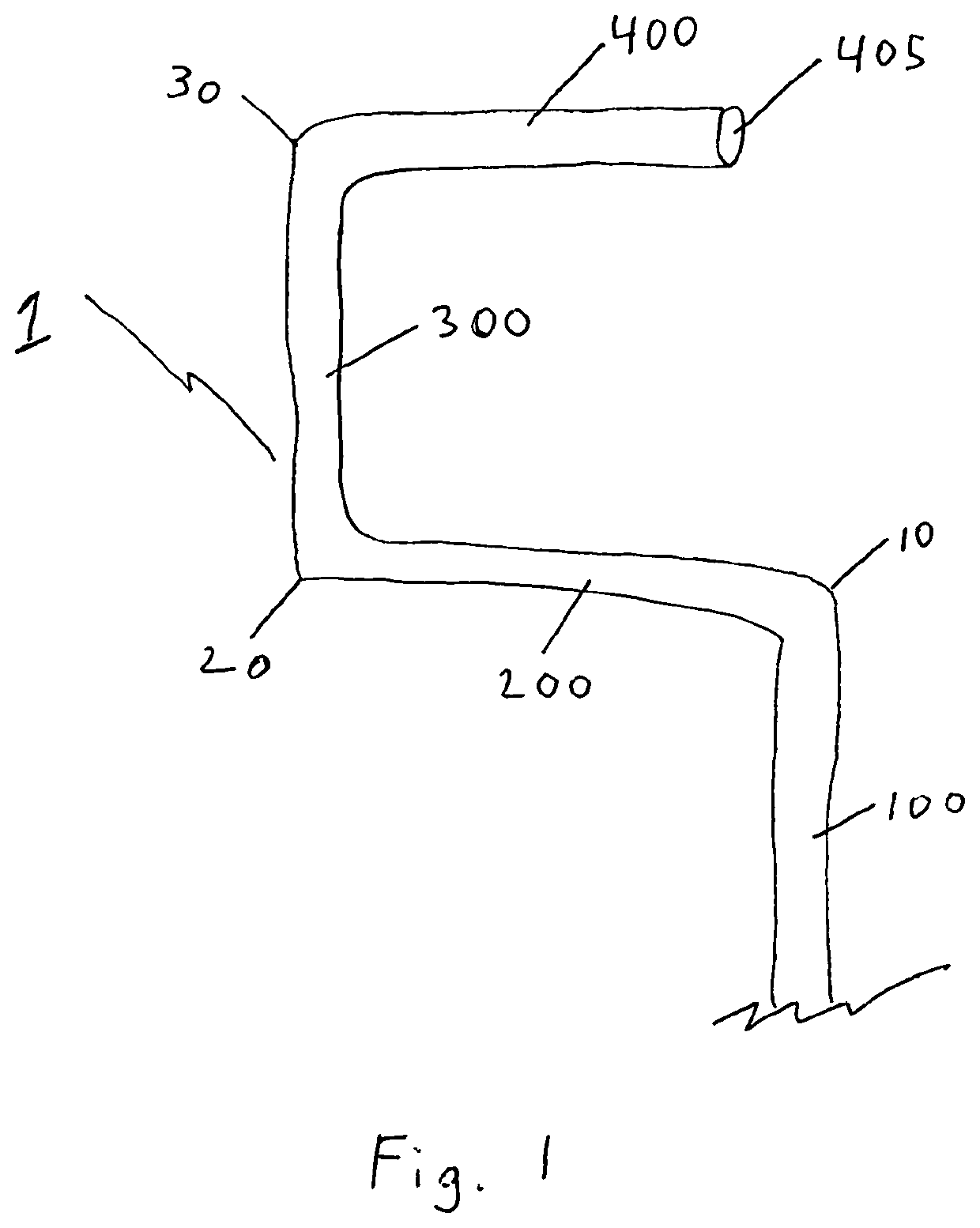
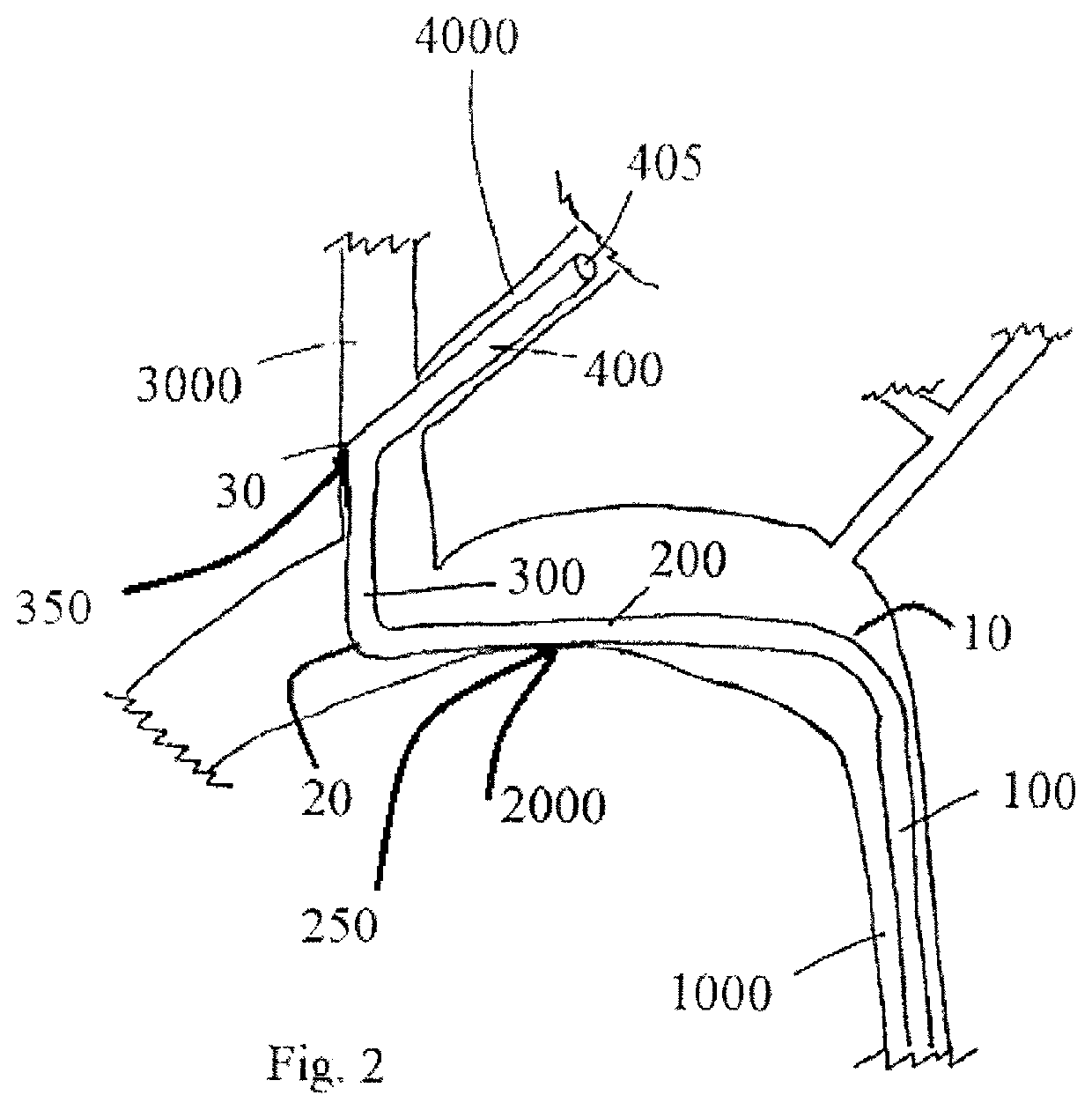