

Medical viewing system and method for detecting boundary structures
- Summary
- Abstract
- Description
- Claims
- Application Information
AI Technical Summary
Benefits of technology
Problems solved by technology
Method used
Image

Examples
Embodiment Construction
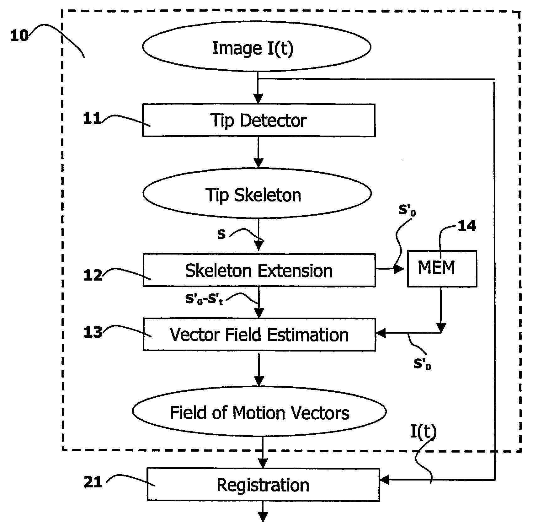
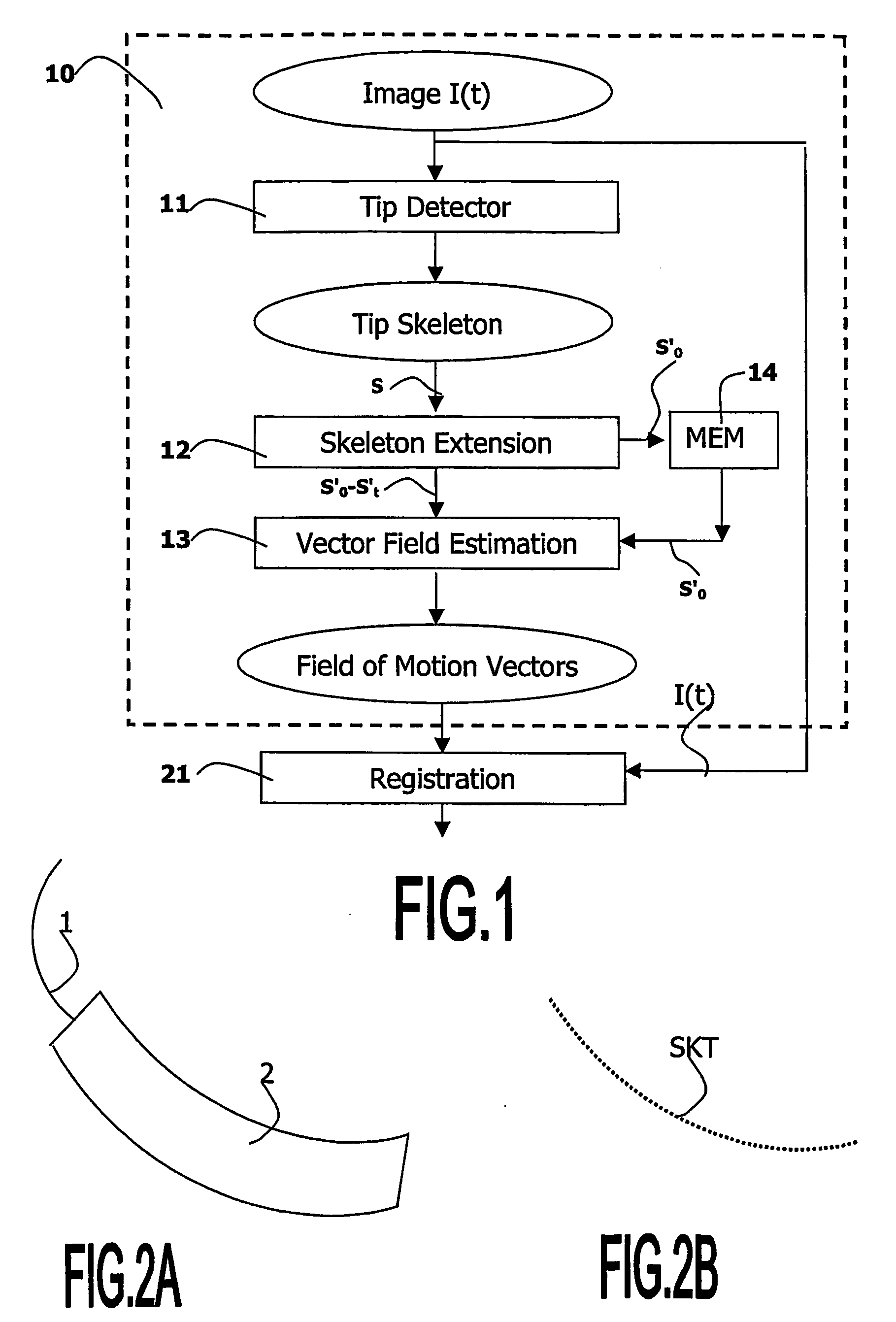
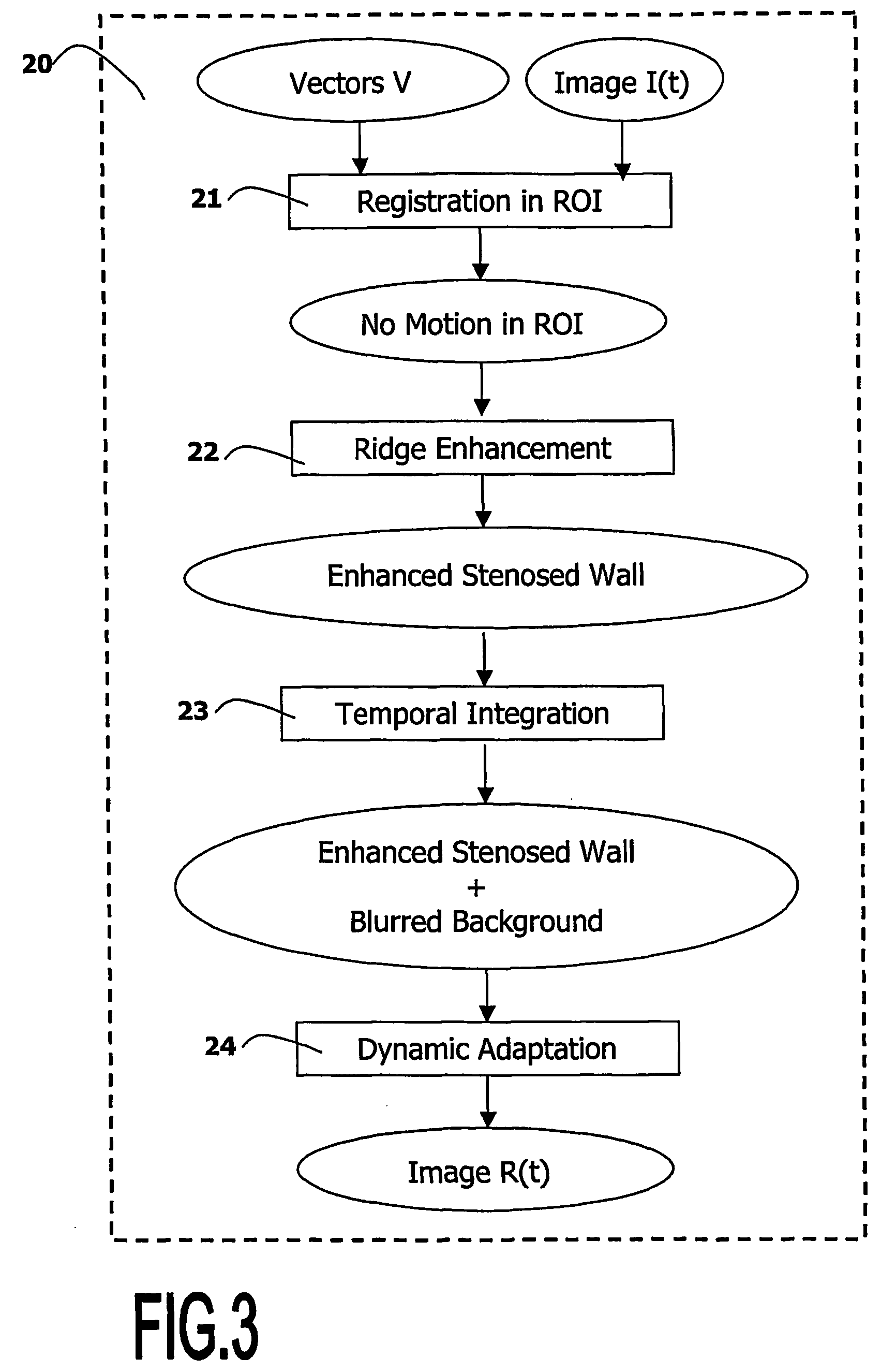
[0020] The invention relates to a viewing system, and to a computer executable image processing method that is used in the viewing system, for detecting, localizing, registering, enhancing and zooming structures in noisy images. The viewing system and the image processing method of the invention are described hereafter in an example of application to the medical field of cardiology. In said application, the object of interest is an artery, which is observed during a medical intervention called angioplasty, in a sequence of X-ray fluoroscopic images called angiograms.
[0021] Referring to FIG. 6A, the angioplasty is a medical intervention that usually comprises several stages for enlarging an artery at the location of a lesion called stenosis. In a preliminary stage, the practitioner localizes a stenosis 3 in a patient's artery 4 as best as possible in peri-interventional images. These images are obtained while using a contrast agent introduced in the artery in order to improve the im...
PUM
 Abstract
Description
Claims
Application Information