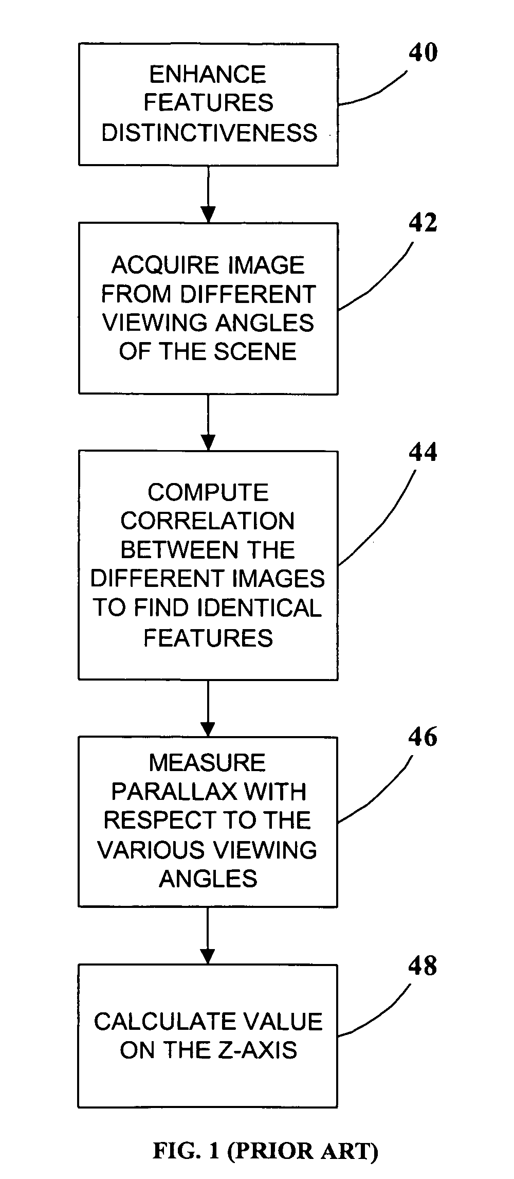
Important constraints in applying medical
optical imaging to the dental field include: space limitations equipment must be compact enough to fit comfortably within the confines of the mouth; time limitations—the image must be formed in a brief time to avoid problems of the movement of the patient as well as the movement of the practitioner and apparatus; and surface detail limitations—accurately plotting 3D surface contours of intra-oral scenes must take into account the absence or paucity of surface detail in many close-up applications, where only partial views of intra-oral features may be available.
In practice, a limitation is imposed by the lack of recognizable surficial surgical features on intra-oral objects.
This limitation is often encountered in situations in which a surface is to be sampled three dimensionally for 3D modeling.
As previously noted, however, a significant limitation in dental work is the lack of surface detail in many situations.
Without a substantial amount of surface detail, it is not possible to unambiguously match surface features for accurate
triangulation.
Unfortunately, however, scanning (which is essentially a one-dimensional, or “1D” approach) can sometimes require excessive time, particularly if
high resolution is desired.
Corby, however, is limited to the use of a one-dimensional scanning methodology, which (as discussed above) may not be suitable for certain dental applications because of the patient / practitioner movement that is likely to be encountered.
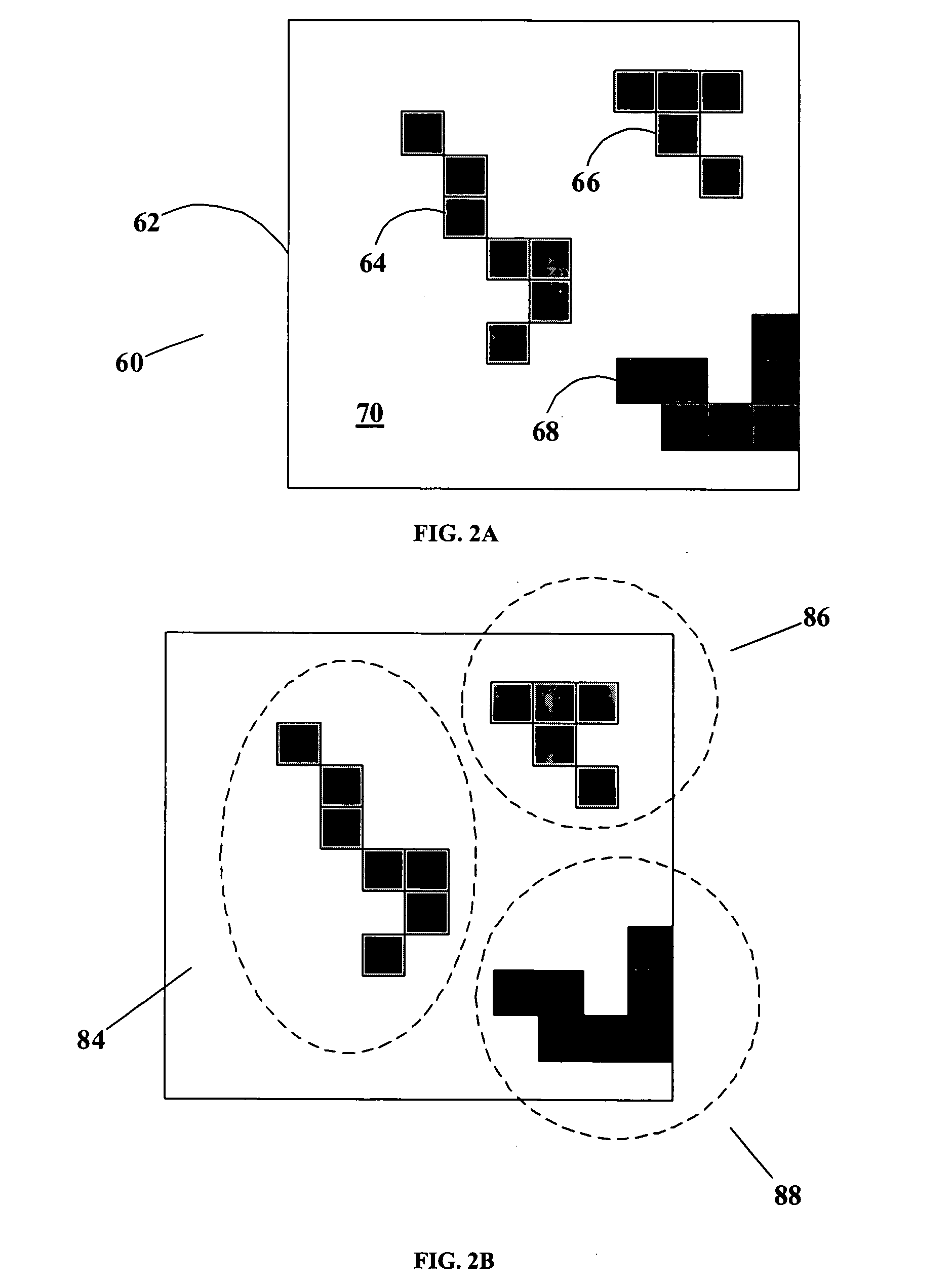
A limitation associated with the employment of regular grids is that of discontinuities and ambiguities.
Broken grid lines are a cause for errors because of the ambiguities introduced by this phenomenon.
A plain point of light is normally indistinguishable from other plain points of light, and so there can arise ambiguities in matching the same point from one image to another.
If two different points are mistakenly matched when triangulating different images, the resulting z-axis calculation will be in error, and the 3D model will be defective.
(The same problem applies to the use of lines.)
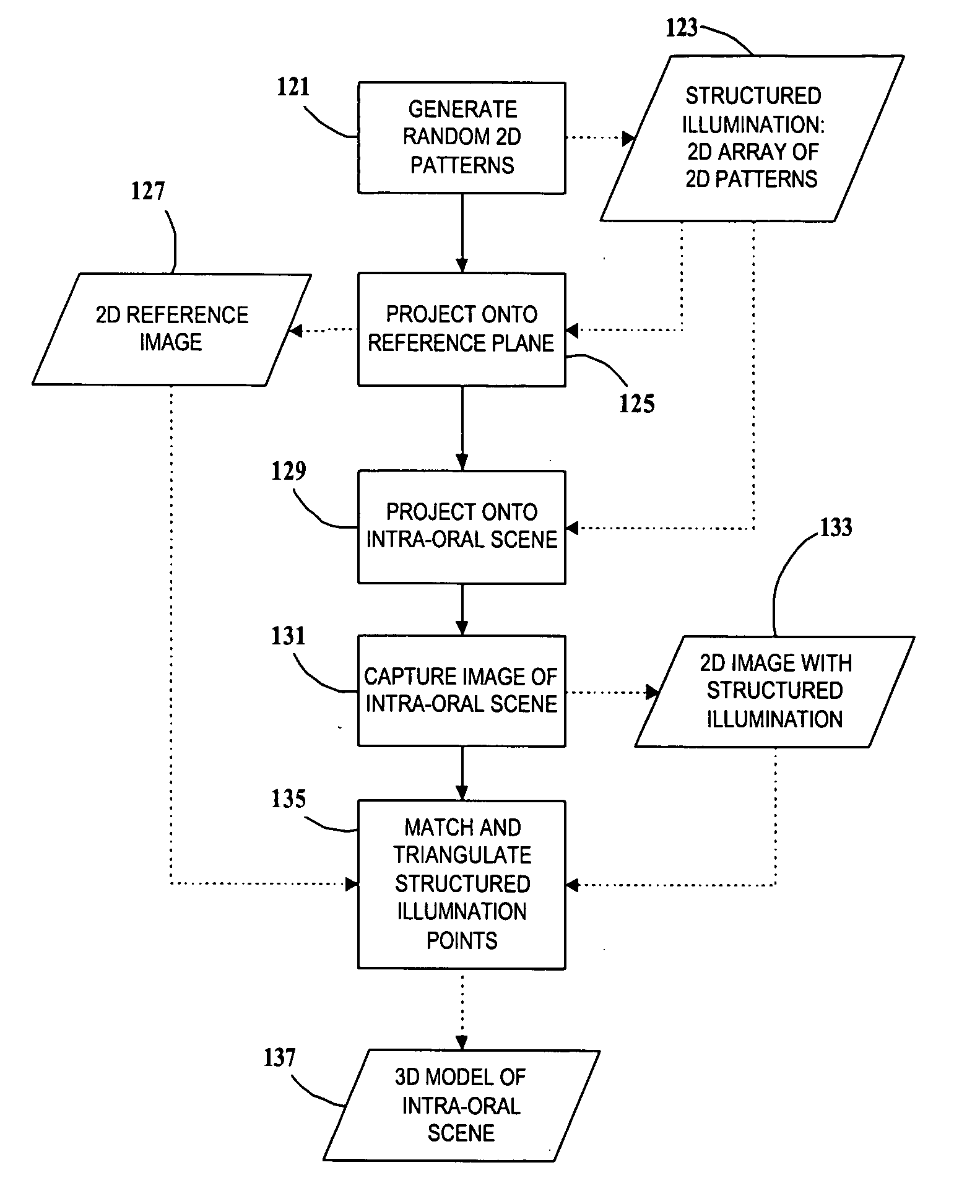
However, this cannot be employed for projected (non-scanned) images or for stored images where features are matched after the structured illumination has been projected, because there is no timing information.
It also cannot be used with the single imaging method mentioned above.
In particular, certain prior art schemes would require illuminating the same areas of the intra-oral scene with patterns over a prolonged time period or repeatedly at different times. These practices, however, can introduce inaccuracies in the measurements due to any relative movement between the dental patient, the apparatus that projects the illumination, and the apparatus that captures the images (camera).
A principal limitation of Albeck '151 is that the random patterns, being created by a physical process (
laser speckle), can be neither stored nor reproduced.
As noted above, an important limitation is imposed by the small space inside the mouth, which does not permit the introduction of bulky apparatus.

