Pre-And Intra-Operative Localization of Penile Sentinel Nodes

Inactive Publication Date: 2008-07-03
NOVADAG TECH INC
View PDF39 Cites 22 Cited by
  • Summary
  • Abstract
  • Description
  • Claims
  • Application Information

AI Technical Summary

Problems solved by technology

One such risk factor is infection with human papillomavirus.
Unfortunately, there is significant anatomical variation in penile lymph node drainage as well as in sentinel lymph node position.
Nevertheless these procedures are difficult, time-consumi

Method used


Image

  • Pre-And Intra-Operative Localization of Penile Sentinel Nodes
  • Pre-And Intra-Operative Localization of Penile Sentinel Nodes
  • Pre-And Intra-Operative Localization of Penile Sentinel Nodes

Examples


example 1

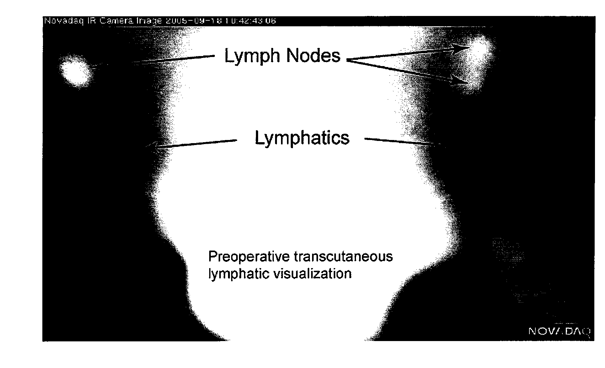
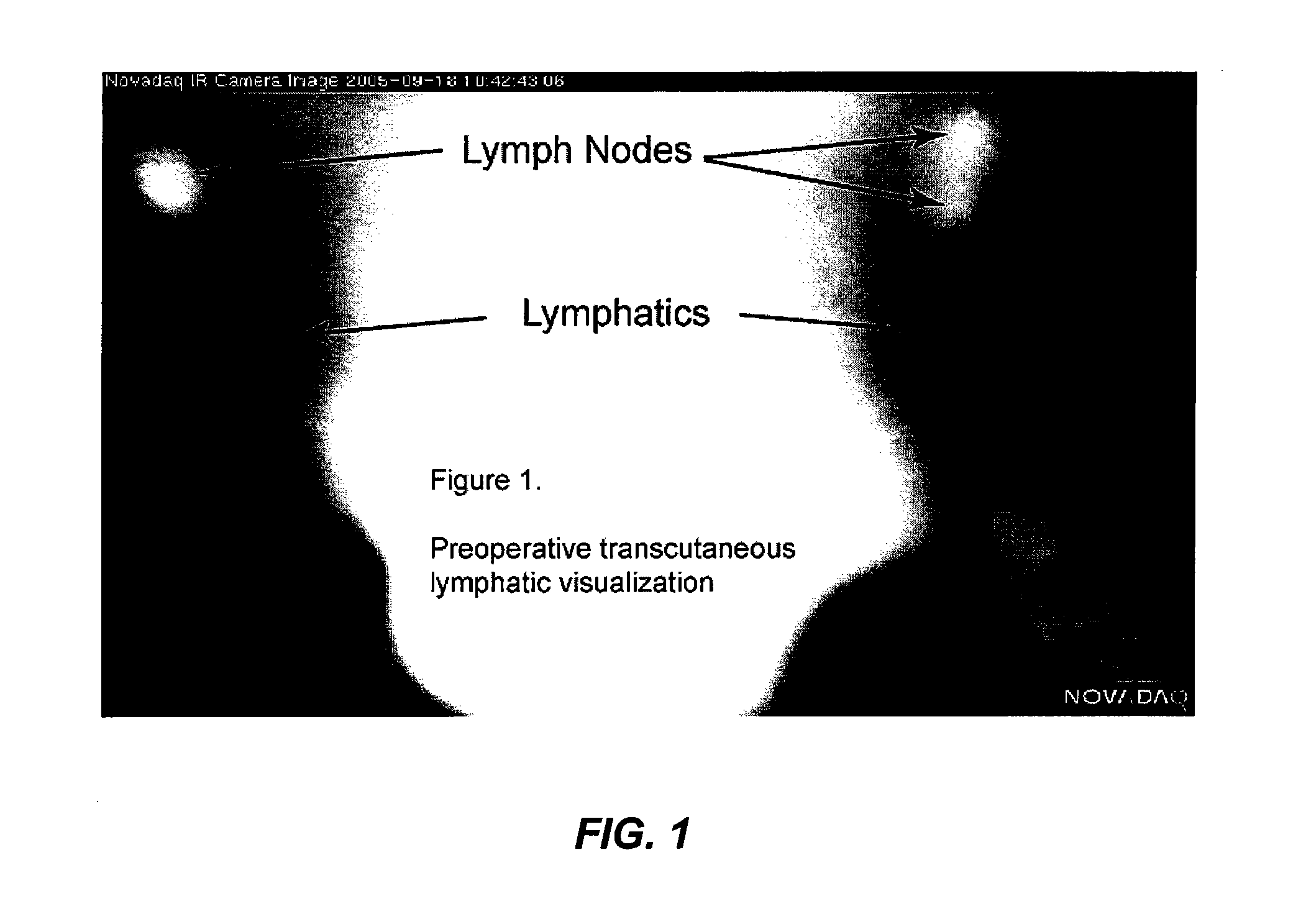
[0041]Intraoperative video angiography is performed with a laser-fluorescence imaging device (Novadaq Technologies, Inc., Mississauga, Ontario, Canada) consisting of a near infrared (NIR) laser light source and a NIR-sensitive digital camcorder. For measurements, the unit is positioned 30 to 40 cm from the area of interest. ICG, dissolved in water, is then injected as a bolus. The NIR light emitted by the laser light source induces ICG fluorescence. The fluorescence is recorded by a digital video camera, with optical filtering to block ambient and laser light so that, when desired, only ICG fluorescence is captured. Images can be observed on screen in real time (25-30 images / sec). The images can be reviewed and stored on the digital video camera or transferred to a computer or to storage media.

example 2

[0042]Sprague-Dawley rats, 60 to 100 days old, weighing 275-325 grams are used. All animals are anesthetized using intraperitoneal injection of Ketamine / Xylazine (40-80 mg / kg and 5 / 10 mg / kg, respectively) or isoflurane. No pre-anesthetic medications are used. When appropriate depth of anesthesia is reached, positioning of the animal takes place. All animals are fastened to a padded and heated restraint device in the supine position using gauze knots to fix all four extremities. Depth of anesthesia, regularity of respirations, and heart beat palpation are repeatedly checked. A pulse oximeter may be used to monitor the animal. Placebo (distilled water) or fluorochromes, ICG or Fluorogold, is administered by intra-penile, sub-albugineal injection of 25 ul of ICG diluted in 100 μl of water for injection, per cavernous body.

[0043]Surgery / Procedure starts after appropriate preparation of surgical field by Povidone-Iodine scrub, 70% Isopropyl Alcohol and Povidone-Iodine solution. The surgi...

example 3

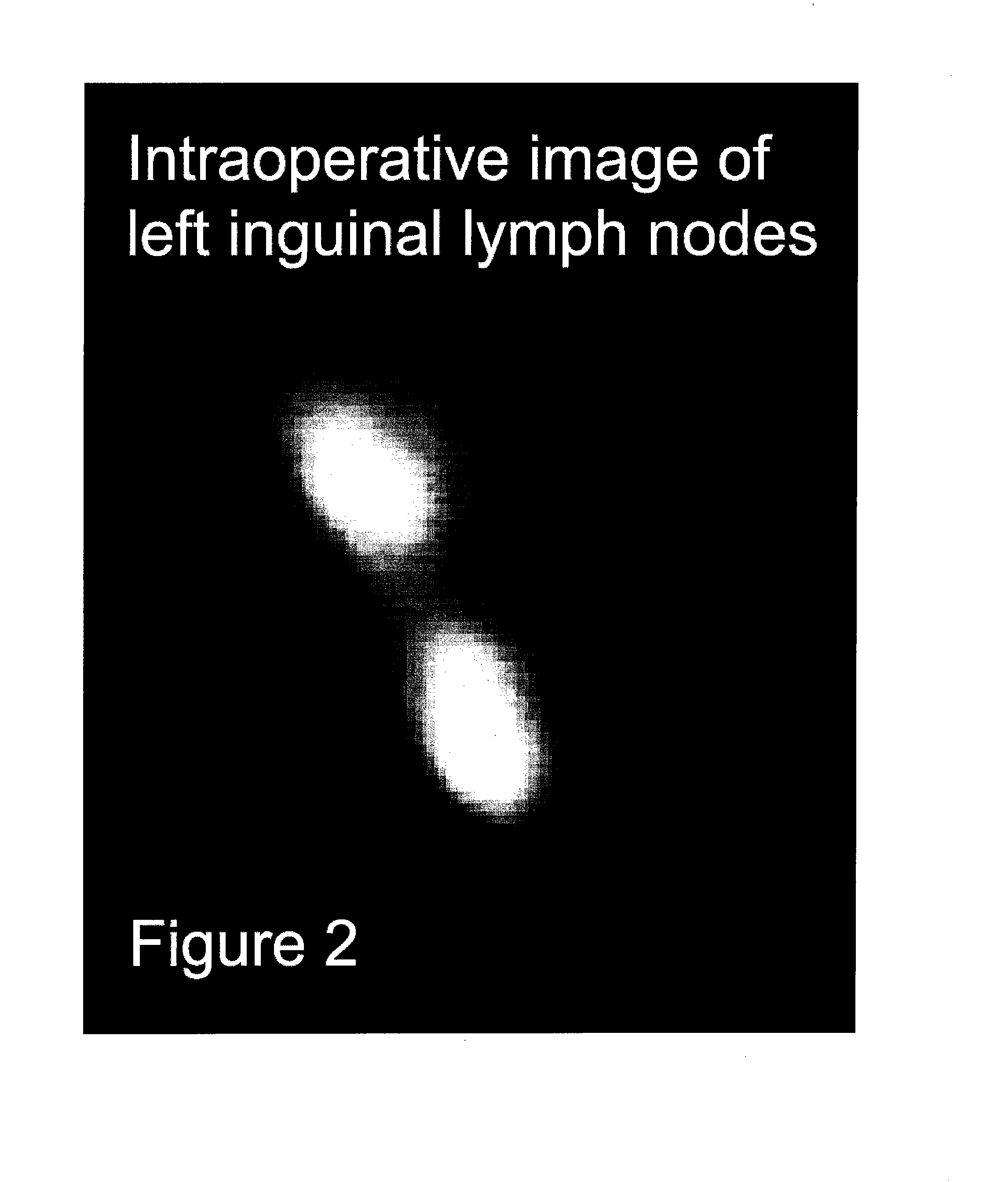
[0047]Twenty two 22 male Sprague-Dawley rats received penile intracavernous ICG injections (20 μl of 0.25 mg / ml). From 2 hours to 7 days later, near infrared fluorescence (NIRF) imaging was performed both before and during dissection. Control rats received comparable saline injections at several time points. A Novadaq intraoperative NIRF imaging system (SPY®) was used for macroscopic fluorescence imaging and an Olympus NIRF microscope IX 70 (Olympus America, Inc., Center Valley, Pa.) was used for histology. All animals were anesthetized and shaved before imaging. All visualized nodes were sent for NIR microscopy and haematoxylin and eosin (H&E) staining for histological examination.


PUM


Abstract

The invention provides methods for pre- and intra-operatively localizing penile sentinel lymph nodes by injection of fluorescent dyes into penile tumors.

Description

CROSS-REFERENCES TO RELATED APPLICATIONS[0001]This disclosure claims the benefit of U.S. Provisional Application No. 60 / 843,175, filed Sep. 7, 2006, the contents of which are incorporated herein by reference.STATEMENT AS TO RIGHTS TO INVENTIONS MADE UNDER FEDERALLY SPONSORED RESEARCH AND DEVELOPMENT[0002]NOT APPLICABLEREFERENCE TO A “SEQUENCE LISTING,” A TABLE, OR A COMPUTER PROGRAM LISTING APPENDIX SUBMITTED ON A COMPACT DISK[0003]NOT APPLICABLEBACKGROUND OF THE INVENTION[0004]A variety of medical techniques suitable for imaging biological tissues and organs are known. These include traditional x-rays, ultra-sound, magnetic resonance imaging, and computerized tomography.[0005]A variety of dyes useful for medical imaging have been described, including radio opaque dyes, fluorescent dyes, and colorimetric dyes (see e.g., U.S. Pat. Nos. 5,699,798; 5,279,298; 6,351,663). Imaging techniques and systems using fluorescent dyes have been described for the heart and eye (see, e.g., U.S. Pat...

Claims


Application Information

Patent Timeline
IPC IPC(8): A61N1/30A61M31/00
CPCA61K49/0034
Inventor GOLIJANIN, DRAGANBRZOZOWSKI, LUKASZWOOD, RONALD
Owner NOVADAG TECH INC
Why Eureka
Try Eureka