Method and System for Model-Based Fusion of Computed Tomography and Non-Contrasted C-Arm Computed Tomography

Active Publication Date: 2013-05-23
SIEMENS HEALTHCARE GMBH
View PDF2 Cites 31 Cited by
  • Summary
  • Abstract
  • Description
  • Claims
  • Application Information

AI Technical Summary

Benefits of technology

[0004]The present invention provides a method and system for fusion of pre-operative and intra-operative imaging modalities. Embodiments of the present invention map high-quality patient-specific models from a pre-operative imaging modality, such as computed tomography (CT), to an intra-operative imaging modality, such as 3D C-arm CT, without adding contrast agent. Embodiments of the present invention use a surrogate anatomy, such as the trachea bifurcation, that is visible in both modalities without adding contrast agent in order to determine an

Problems solved by technology

However, such intra-operative imaging modalities typically have limited image quality of soft tissue and reliable assessment of the cardiac

Method used


Image

  • Method and System for Model-Based Fusion of Computed Tomography and Non-Contrasted C-Arm Computed Tomography
  • Method and System for Model-Based Fusion of Computed Tomography and Non-Contrasted C-Arm Computed Tomography
  • Method and System for Model-Based Fusion of Computed Tomography and Non-Contrasted C-Arm Computed Tomography

Examples


Example

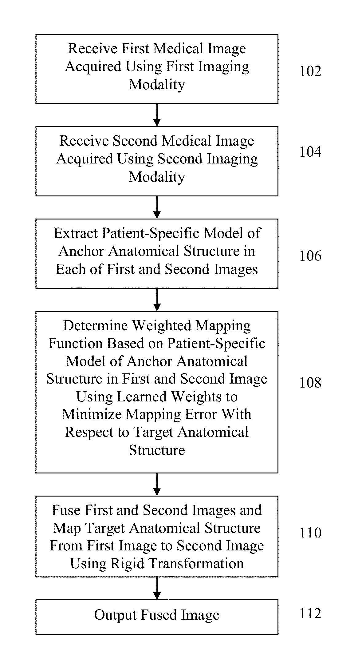
[0014]The present invention relates to model-based fusion of multi-modal volumetric images. Embodiments of the present invention can be used to fuse image information in multiple imaging modalities, such as computed tomography (CT), Dyna CT, echocardiography data, fluoroscopic image data, and magnetic resonance imaging (MRI). Embodiments of the present invention are described herein to give a visual understanding of the model-based image fusion method. A digital image is often composed of digital representations of one or more objects (or shapes). The digital representation of an object is often described herein in terms of identifying and manipulating the objects. Such manipulations are virtual manipulations accomplished in the memory or other circuitry / hardware of a computer system. Accordingly, is to be understood that embodiments of the present invention may be performed within a computer system using data stored within the computer system.

[0015]Embodiments of the present inve...


PUM


Abstract

A method and system for model-based fusion of multi-modal volumetric images is disclosed. A first patient-specific model of an anchor anatomical structure is detected in a first medical image acquired using a first imaging modality, and a second patient-specific model of the anchor anatomical structure is detected in a second medical image acquired using a second imaging modality. A weighted mapping function is determined based on the first patient-specific model of the anchor anatomical structure and the second patient-specific model of the anchor anatomical structure using learned weights to minimize mapping error with respect to a target anatomical structure. The target anatomical structure from the first medical image to the second medical image using the weighted mapping function. In an application of this model-based fusion to transcatheter valve therapies, the trachea bifurcation is used as the anchor anatomical structure and the aortic valve is the target anatomical structure.

Description

[0001]This application claims the benefit of U.S. Provisional Application No. 61 / 563,082, filed Nov. 23, 2011, the disclosure of which is herein incorporated by reference.BACKGROUND OF THE INVENTION[0002]The present invention relates to fusion of multi-modal volumetric medical images, and more particularly, to model-based fusion of pre-operative computed tomography (CT) and intra-operative non-contrasted C-arm CT images.[0003]In recent years, there has been a major trend in cardiac therapy towards minimally invasive procedures to reduce the risks involved with classical surgical techniques. Transcatheter valve interventions are becoming the standard therapy for high risk surgical patients. Such procedures off the potential to reduce morbidity, mortality, and costs of surgical valve replacement or repair, while accelerating patient recovery. One type of minimally invasive procedure is Transcatheter Aortic Valve Replacement (TAVI). The TAVI procedure involves accessing a femoral arter...

Claims


Application Information

Patent Timeline
IPC IPC(8): G06T7/00
CPCG06T7/0012G06T7/0032G06T2200/04G06T2207/10081G06T2207/30101G06T2207/20081G06T2207/20221G06T2207/30048G06T2207/30061G06T2207/10121G06T7/344
Inventor GRBIC, SASAIONASEC, RAZVAN IOANJOHN, MATTHIASBOESE, JANGESELL, CHRISTIANCOMANICIU, DORIN
Owner SIEMENS HEALTHCARE GMBH
Why Eureka
Try Eureka