Methods and systems for determining a transformation function to automatically register different modality medical images

a technology of transformation function and medical image, applied in the field of medical imaging system, can solve the problems of requiring a significant amount of time and/or computing resources for registration and segmentation operations

Active Publication Date: 2014-01-30
GENERAL ELECTRIC CO
View PDF3 Cites 43 Cited by
  • Summary
  • Abstract
  • Description
  • Claims
  • Application Information

AI Technical Summary

Benefits of technology

The patent describes a method for creating a registered organ model and using it to determine the position of an organ within a specific area. This information can be used to identify different types of lesions or to segment the organ for further analysis. The technical effect of this method is to provide a more accurate and reliable way to analyze and diagnose organ abnormalities.

Problems solved by technology

The patent text discusses the challenges of registering and segmenting medical images, which can be time-consuming and resource-intensive. Additionally, a large number of clinical applications require specific information to facilitate these operations. The technical problem addressed by the patent is to provide a more efficient and automated method for registering and segmenting medical images.

Method used


Image

  • Methods and systems for determining a transformation function to automatically register different modality medical images
  • Methods and systems for determining a transformation function to automatically register different modality medical images
  • Methods and systems for determining a transformation function to automatically register different modality medical images

Examples


Embodiment Construction

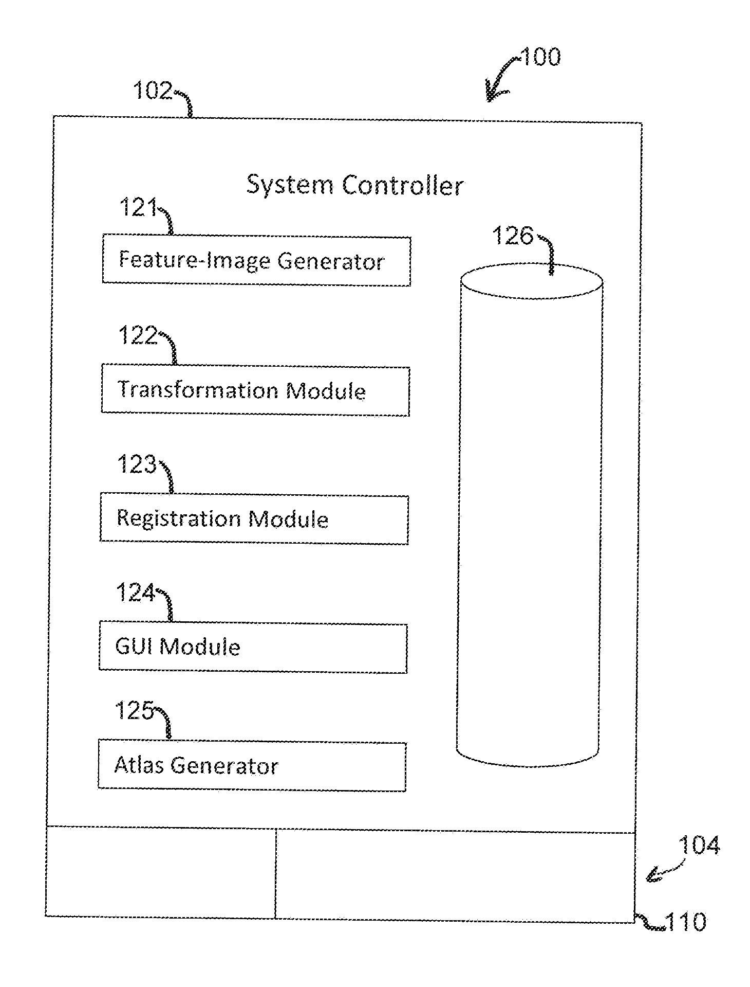
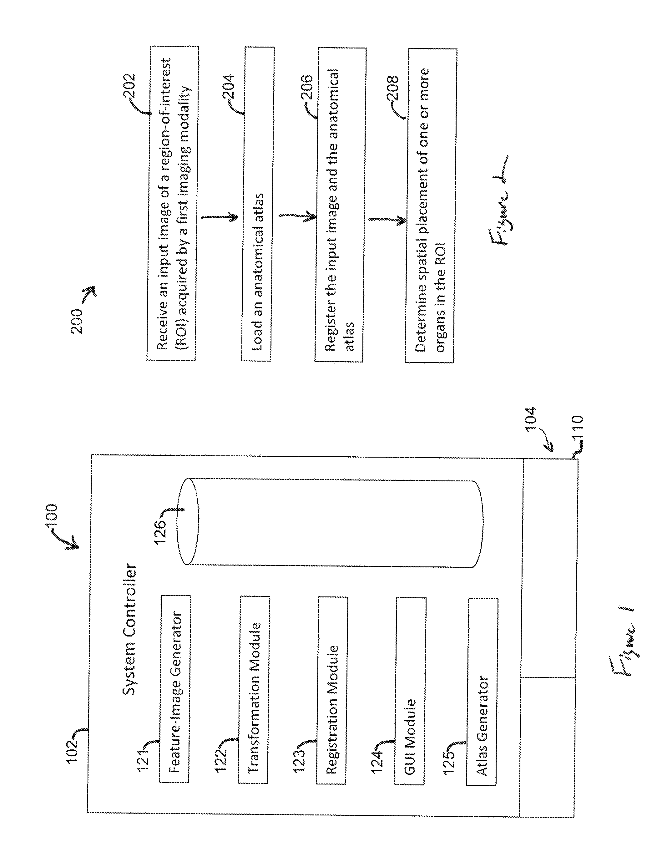
[0016]Embodiments described herein include methods, systems, and computer readable media that may facilitate at least one of processing or analyzing medical images of a region-of-interest (ROI) (also referred to as a volume-of-interest (VOI)). For example, embodiments may include methods, systems, and computer readable media that generate a multi-modality anatomical atlas. Embodiments may also include methods, systems, and computer readable media that determine a spatial placement of one or more organs in a region-of-interest (ROI).

[0017]The medical images may include image data or datasets that represent a visualization of the ROI. The image data may include pixels (or voxels) having signal intensity values or other values / qualities / characteristics that may be processed to form the visualization. Various imaging modalities may be used to acquire the medical images. Non-limiting examples of such modalities include ultrasound, magnetic resonance imaging (MRI), computed tomography (CT...


PUM


Abstract

A method that includes receiving an input image of a region of interest (ROI) of an individual. The input image is a medical image acquired by a first imaging modality. The method also includes generating a first feature image based on the input image. The first feature image includes a designated anatomical feature of the ROI. The method also includes obtaining an anatomical atlas. The atlas has a reference image of the ROI of at least one other individual and an organ model. The reference image is a medical image that is acquired by a second imaging modality that is different from the first imaging modality. The method also includes determining a transformation function by registering the first feature image with a second feature image that is based on the reference image and includes the designated anatomical feature.

Description


Claims


Application Information

Patent Timeline
Owner GENERAL ELECTRIC CO
Why Eureka
Try Eureka