Diagnostic biomarkers and therapeutic targets for pancreatic cancer

a pancreatic cancer and biomarker technology, applied in the field of cancer diagnostics, prognostics, and therapeutics, can solve the problems of low survival statistics, limited finding of t cell antigens, ineffective treatment, etc., and achieve the effect of long-term disease-free survival

Inactive Publication Date: 2016-11-10
THE JOHN HOPKINS UNIV SCHOOL OF MEDICINE
View PDF0 Cites 1 Cited by
  • Summary
  • Abstract
  • Description
  • Claims
  • Application Information

AI Technical Summary

Benefits of technology

[0011]According to another embodiment a method predicts response to a pancreatic cancer vaccine in a human. A body sample of the human is contacted with at least one antibody that specifically binds to a protein selected from the group consisting of: Transferrin receptor (TFRC), regulatory subunit 12A of protein phosphatase 1 (PPP1R12A), and regulatory subunit 8 of the 26S proteasome (PSMC5). The amount of antigen bound to the antibody is detected. A decreased amount of antigen bound to the antibody relative to an amount bound to a control sample prior to vaccination predicts long term disease-free survival.
[0013]According to another embodiment a method predicts response to a pancreatic cancer vaccine in a human A sample of the human comprising antibodies is contacted with at least one protein selected from the group consisting of: Transferrin receptor (TFRC), regulatory subunit 12A of protein phosphatase 1 (PPP1R12A), and regulatory subunit 8 of the 26S proteasome (PSMC5). The amount of antibody bound to the at least one protein is detected. An increased amount of antibody bound to the at least one protein relative to an amount bound to a control sample obtained prior to vaccination predicts long term disease-free survival.

Problems solved by technology

Inadequate early diagnosis, resistance to current therapies, and ineffective treatment account for these low survival statistics.
However, finding T cell antigens is limited by the need for large amounts of patient lymphocytes and the lack of reagents for each patient-specific HLA (6).
However, this process has an inherent bias towards identifying proteins that are abundantly expressed (11).
Therefore, this process is inadequate for detecting membrane-associated proteins, the most relevant category of proteins as potential biomarkers.
This process is inefficient in separating single proteins, which obscures which protein instigates the antibody response.
Furthermore, low abundant antigens are generally overshadowed by high abundant proteins with the same molecular weight in this process.
However, these arrays do not identify which specific protein in the fraction instigates the immune response and there also issues with fractionation.
The protein arrays utilizing printed monoclonal antibodies are potentially limited by reagent availability thereby preventing an unbiased proteome being used because a high affinity and highly specific monoclonal antibody is needed for each protein to be probed.

Method used


Image

  • Diagnostic biomarkers and therapeutic targets for pancreatic cancer
  • Diagnostic biomarkers and therapeutic targets for pancreatic cancer
  • Diagnostic biomarkers and therapeutic targets for pancreatic cancer

Examples


example 1

Materials and Methods

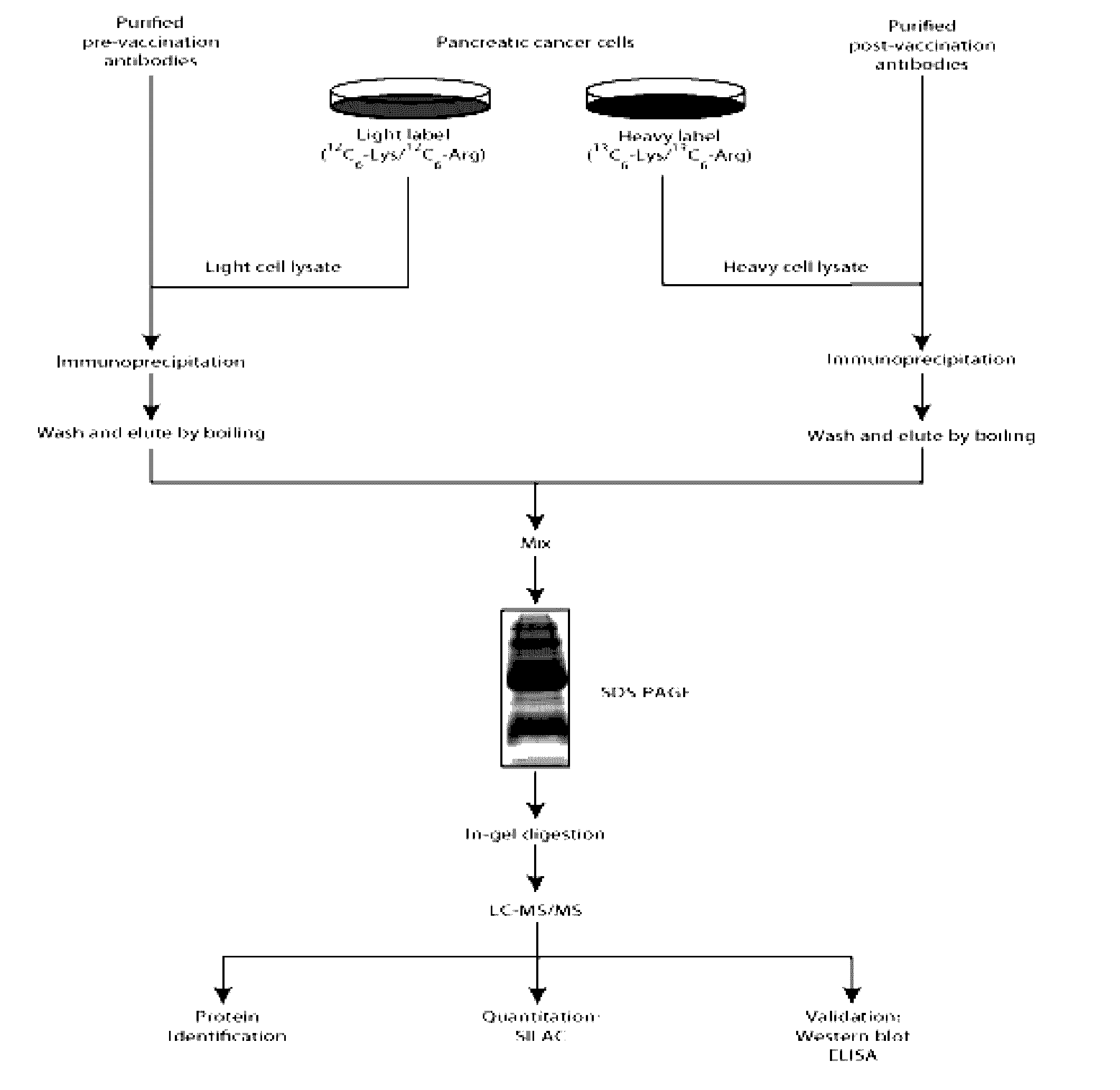
Patients, Serum and Tissue Samples

[0044]Patients were enrolled in a phase II study of an allogeneic GM-CSF secreting whole cell pancreatic cancer vaccine in compliance with the Johns Hopkins Medical Institution Institutional Review Board (IRB)-approved J9988 protocol. Blood samples were collected pre-vaccination, 14 days after 1St vaccination and 28 days after each subsequent vaccination. Sera was collected by centrifugation, aliquoted and stored at −80° C. Pancreatic tumor tissue samples were obtained from patients prior to vaccination.

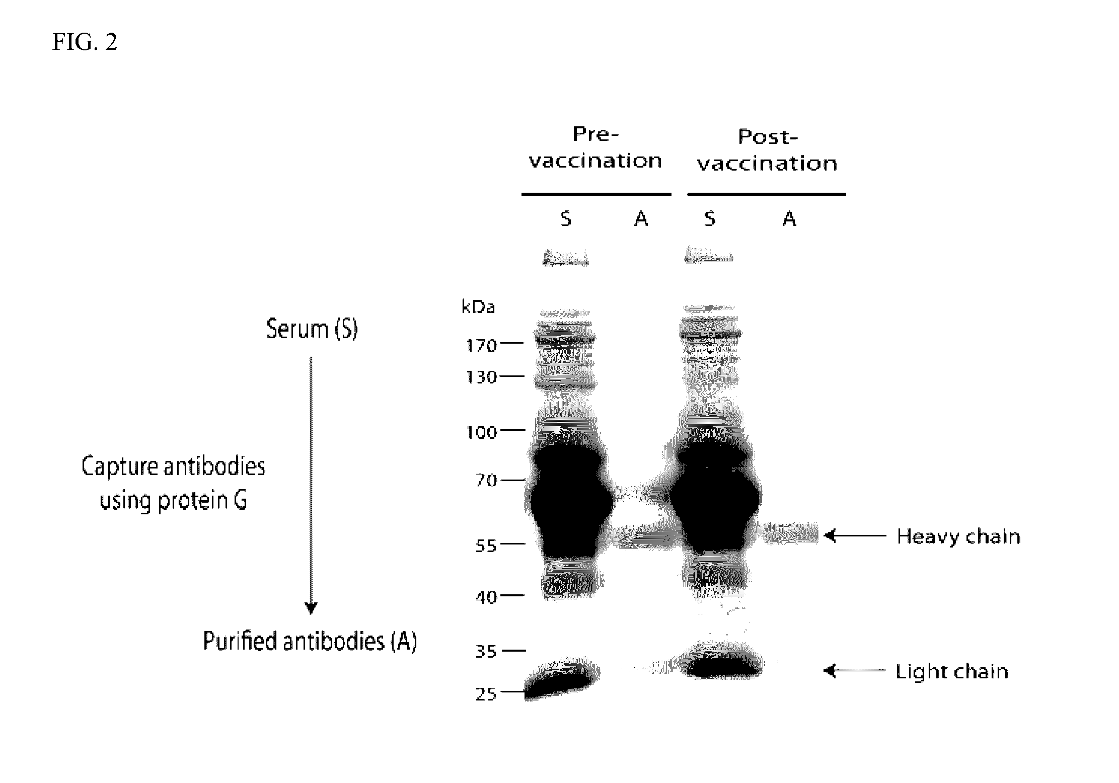
Antibody Purification

[0045]Antibodies were purified from pre- and post-3rd vaccination sera using a protein G column (GE Healthcare, Piscataway, N.J., USA) as per manufacturer's protocol. Quantification of purified antibodies was done using NanoDrop spectrophotometer (Thermo Fisher Scientific, Waltham, Mass., USA).

Sample Preparation

[0046]The human pancreatic cancer cell line, Panc 10.05 was grown as previously described. For the ...

example 2

Design and Validation of Quantitative Proteomic Approach

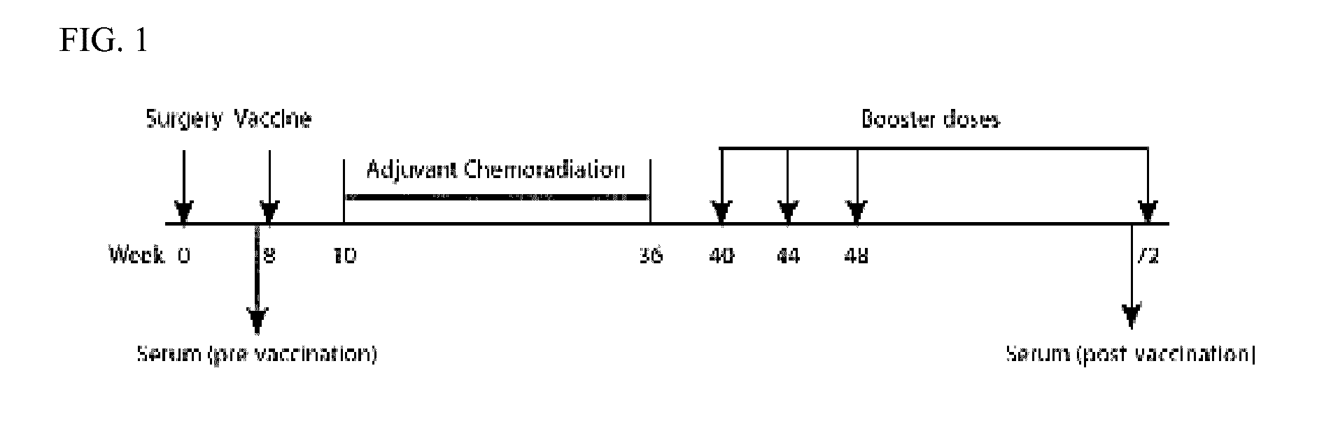
[0052]60 pancreatic cancer patients, who had their pancreas surgically removed, were involved in the study (FIG. 1) (2). The patients received their first vaccination 2 months after surgery. One month after the first vaccination, the patients underwent a 6-month course of chemoradiation. The second, third and fourth vaccines were each administered at sequential one-month intervals from the time of chemotherapy completion. The fifth, and final, vaccination was received 6 months after the fourth vaccination. Serum samples were obtained pre- and post-vaccination for all five vaccinations (2). The 60 vaccinated patients were divided into 3 groups (A, B and C) based on length of disease free survival (DFS) (2). Group A was composed of 12 patients who received all of the scheduled vaccinations and demonstrated a DFS>3 years (prolonged DFS as well as overall survival). The clinical time point cutoff was determined to be 3 years becaus...

example 3

Identification of Proteins by the SASI Approach

[0053]To identify the proteins in the post-vaccination sera of patients in Group A (DFS>3 years), we used the immunized sera from three patients (patients 9, 27 and 52) who demonstrated other evidence of post-vaccination immune responses. We identified a total of 976 proteins for patient 9, 811 proteins for patient 27 and 727 proteins for patient 52 (FIG. 5). A broad range of post-vaccination antibody response was observed; from a 16 fold change increase post-vaccination to a 10 fold change decrease. The majority of the proteins, as expected, had no change in response post-vaccination. We identified 51 proteins for patient 9, 47 proteins for patient 27 and 54 proteins for patient 52 that had a 2 fold change in response. Through the SASI approach, we present the first large scale study to identify and categorize proteins that are targeted by antibodies in the human body.

[0054]Pre-vaccination and post-4th vaccination sera from 3 patients,...


PUM

PropertyMeasurementUnit
mass toleranceaaaaaaaaaa
temperatureaaaaaaaaaa
thickaaaaaaaaaa

Abstract

We identified >40 proteins that elicited at least a 2-fold increase in antibody response post-pancreatic-cancer vaccination, from each of three patients' sera. The antibody responses detected against these proteins in patients with >3 years disease-free survival indicates the anti-tumor potential of targeting these proteins. We found that tissue expression of proteins PSMC5, TFRC and PPP1R12A increases during tumor development from normal to pre-malignant to pancreatic tumor. In addition, these proteins were shown to be pancreatic cancer-associated antigens that are recognized by post-vaccination antibodies in the sera of patients that received the vaccine and have demonstrated a favorable disease free survival.

Description

CROSS REFERENCE TO RELATED APPLICATIONS[0001]This application is a continuation of U.S. application Ser. No. 14 / 649,248, filed on Jun. 3, 2015, which is a national stage entry of International Application No. PCT / US2013 / 072592, filed on Dec. 2, 2013, which claims priority to U.S. Provisional Patent Application No. 61 / 732,402, filed on Dec. 3, 2012, each of which is incorporated herein by reference in its entirety.FEDERALLY SPONSORED RESEARCH OR DEVELOPMENT[0002]This invention was made with government support under Grant No. P50CA62924 awarded by the National Institutes of Health / National Cancer Institute. The government has certain rights in the invention.TECHNICAL FIELD OF THE INVENTION[0003]This invention is related to the area of cancer diagnostics, prognostics, and therapeutics. Moreover, it relates to the area of immunotherapeutics.BACKGROUND OF THE INVENTION[0004]Pancreatic ductal adenocarcinoma is the fourth leading cause of cancer-related death in the U.S. (1). It is notably...

Claims


Application Information

Patent Timeline
Patent Type & Authority Applications(United States)
IPC IPC(8): G01N33/574
CPCG01N33/57438G01N2333/4703G01N2333/79G01N2333/705G01N2333/916
Inventor JAFFEE, ELIZABETH A.JHAVERI, DARSHIL T.ANDERS, ROBERT
Owner THE JOHN HOPKINS UNIV SCHOOL OF MEDICINE
Why Eureka
Try Eureka