Extrapleural airway device and method

a technology of gaseous exchange and lung, applied in the field of improving the gaseous exchange in the lung of an individual, can solve the problems of copd-related muscle weakness, inability to perform common daily activities, and little hope of recovery, and achieve the effect of improving the gaseous exchange in the lung

Active Publication Date: 2008-09-09
BRONCUS MEDICAL
View PDF17 Cites 68 Cited by
  • Summary
  • Abstract
  • Description
  • Claims
  • Application Information

AI Technical Summary

Benefits of technology

[0017]The devices and methods described herein serve to improve the gaseous exchange in the lungs. In one variation of the present invention, an extrapleural or extraparenchymal airway such as the trachea is fluidly connected to the lung with a conduit. The conduit includes a first end portion, a second end portion and a passageway extending between the end portions. The end portions are adapted to secure the conduit to the tissue structures such that the extrapleural airway is in direct fluid communication with the lung.
[0021]The inventive method may also include the step of delivering a sealant with the conduit. The sealant may be disposed on the exterior of the conduit. The sealant may also be delivered separate from the conduit. The sealant serves to secure the conduit in place, hold the tissues together, prevent side flow around the conduit, and perhaps affect the wound healing response of the tissue to decrease the likelihood that the conduit will be ejected.

Problems solved by technology

Those inflicted with COPD face disabilities due to the limited pulmonary functions.
Usually, individuals afflicted by COPD also face loss in muscle strength and an inability to perform common daily activities.
Since the damage to the lungs is irreversible, there is little hope of recovery.
Most times, the physician cannot reverse the effects of the disease but can only offer treatment and advice to halt the progression of the disease.
However, these conducting airways do not take part in gas exchange because they do not contain alveoli.
As a result, the lungs cannot evacuate all of the inspired air.
The destruction of the alveolar walls results in a dual problem of reduction of elastic recoil and the loss of tethering of the airways.
Unfortunately for the individual suffering from emphysema, these two problems combine to result in extreme hyperinflation (air trapping) of the lung and an inability of the person to exhale.
In this situation, the individual will be debilitated since the lungs are unable to perform gas exchange at a satisfactory rate.
While alveolar wall destruction decreases resistance to collateral ventilation, the resulting increased collateral ventilation does not benefit the individual since air is still unable to flow into and out of the lungs.
Yet, bronchodilator drugs are only effective for a short period of time and require repeated application.
Moreover, the bronchodilator drugs are only effective in a certain percentage of the population of those diagnosed with COPD.
Unfortunately, aside from the impracticalities of needing to maintain and transport a source of oxygen for everyday activities, the oxygen is only partially functional and does not eliminate the effects of the COPD.
Moreover, patients requiring a supplemental source of oxygen are usually never able to return to functioning without the oxygen.
However, lung reduction surgery is an extremely traumatic procedure which involves opening the chest and thoracic cavity to remove a portion of the lung.
If the entire lung is emphysematous, however, removal of a portion of the lung removes gas exchanging alveolar surfaces, reducing the overall efficiency of the lung.
Lung volume reduction surgery is thus not a practical solution for treatment of emphysema where the entire lung is diseased.
Both bronchodilator drugs and lung reduction surgery fail to capitalize on the increased collateral ventilation taking place in the diseased lung.

Method used


Image

  • Extrapleural airway device and method
  • Extrapleural airway device and method
  • Extrapleural airway device and method

Examples


Embodiment Construction

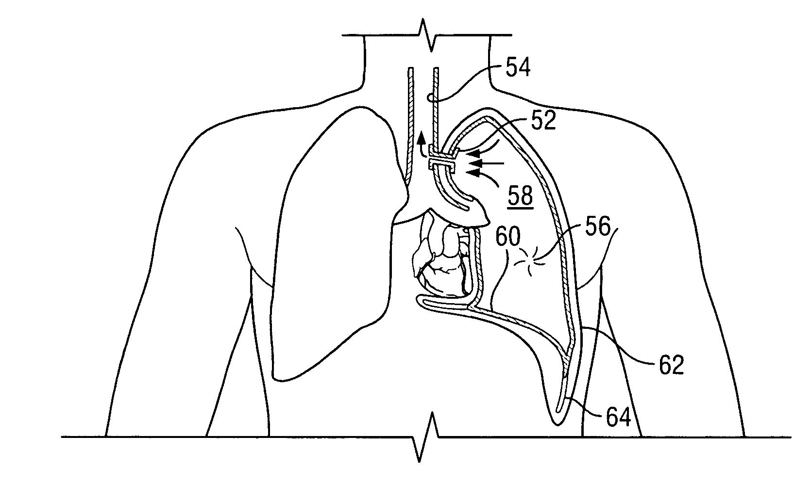
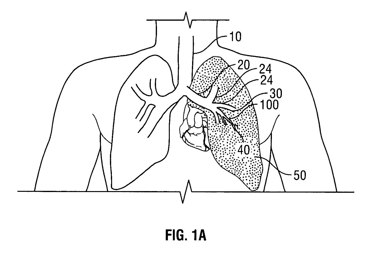
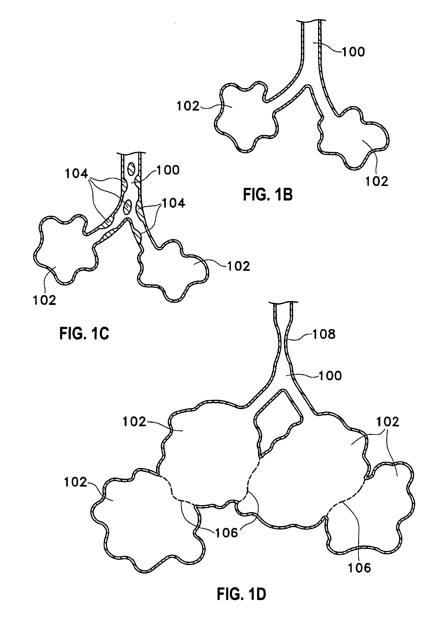
[0042]Described herein are methods and devices for improving the gaseous exchange in a lung. More particularly, a method is described for improving the gaseous exchange in a lung of an individual having chronic obstructive pulmonary disease. The inventive method generally includes fluidly connecting the lung with an extrapleural airway such as the trachea using a conduit. Once the tissue structures are in fluid communication, gas may flow or pass directly from the parenchymal tissue of the lung to the extrapleural airway such as the trachea via the deployed conduit. By “pass directly” from the lung to the extrapleural airway it is meant that at least some volume of gas flows direct from the inner tissue of the lung to the extrapleural airway without passing through typical flow pathways such as the bronchioles, bronchus, etc. which may be constricted or otherwise flow-resistant. Also, by “extrapleural airway” it is meant any airway or portion of an airway that is outside of the pleu...


PUM


Abstract

This is directed to improving the gaseous exchange in a lung of an individual and particularly, this is directed to improving the gaseous exchange in individuals having chronic obstructive pulmonary disease. It generally includes fluidly connecting the lung to an extrapleural airway such as the trachea. In one variation, a conduit is deployed to place the lung and the trachea in fluid communication which allows trapped oxygen-reduced air to pass directly out of the lung and into the trachea. Removing nonfunctional air from the lung tends to improve the gaseous exchange of oxygen into the blood and decompress hyper-inflated lungs. Sealant and biocompatible adhesives may be provided on the exterior of the conduit to prevent side flow, leaks and to otherwise prevent air from entering spaces not intended to receive air such as the pleura space.

Description

REFERENCE TO PRIOR APPLICATIONS[0001]This application claims benefit of 60 / 393,964 Jul. 5, 2002.FIELD OF THE INVENTION[0002]This is directed to improving the gaseous exchange in a lung of an individual and more particularly, this is directed to improving the gaseous exchange in a lung of an individual having chronic obstructive pulmonary disease.BACKGROUND OF THE INVENTION[0003]In 1995, the American Lung Association (ALA) estimated that between 15-16 million Americans suffered from chronic obstructive pulmonary disease (COPD) which includes diseases such as chronic bronchitis, emphysema, and some types of asthma. The ALA estimated that COPD was the fourth-ranking cause of death in the U.S. The ALA estimates that the rates of emphysema is 7.6 per thousand population, and the rate for chronic bronchitis is 55.7 per thousand population.[0004]Those inflicted with COPD face disabilities due to the limited pulmonary functions. Usually, individuals afflicted by COPD also face loss in muscl...

Claims


Application Information

Patent Timeline
Patent Type & Authority Patents(United States)
IPC IPC(8): A61B18/18A61F2/04
CPCA61F2/04A61F2002/046
Inventor LOOMAS, BRYANKAPLAN, GARY
Owner BRONCUS MEDICAL
Why Eureka
Try Eureka