Quantitative single image-based magnetization transfer weighted imaging using an inter-subject normalization reference within the image

a single image and weighted imaging technology, applied in the field of magnetic resonance imaging, can solve the problems of limiting the quality of computed mtr, uncertainty in correspondence, and inability to quantitatively track the progression of disease for a given patient with mtw imaging alone, and achieve the effect of early quantification

Active Publication Date: 2010-03-09
THE JOHN HOPKINS UNIV SCHOOL OF MEDICINE
View PDF3 Cites 7 Cited by
  • Summary
  • Abstract
  • Description
  • Claims
  • Application Information

AI Technical Summary

Benefits of technology

[0019]An advantage of the present invention is that it provides a more precise visualization and quantitative assessment of loss of solid components within white matter.
[0020]Another advantage of the present invention is that it better enables early quantification of myelin loss in several white matter diseases in the spinal cord and can thus follow disease progression at an early stage of the disease.

Problems solved by technology

First, motion induced errors between the reference scan and the saturation scan limit the quality of the computed MTR.
Second, coil loading effects of the MR scanner affect the repeatability of typical MTw images for a given patient, therefore, while the extent of demyelination may be determined for a given patient at a single time point using MTw imaging, it is generally not possible to quantitatively track the progression of the disease for a given patient with MTw imaging alone.
Motion control-induced errors result from the fact that Mz(ω) and Mo are taken from two different images that are acquired at different times. Since these two signals are acquired during two different scans that are temporally separated, the voxels must be co-registered, which results in uncertainty in their correspondence if the co-registration is not perfect.
Any motion of the target tissue between these scans reduces the precision of the Magnetization Transfer Ratio, and thus decreases the Signal to Noise Ratio (SNR) of the resultant MT image and dilutes the quantitative assessment of the tissue MT effect.
Out of plane motion between image acquisitions is particularly problematic in that image manipulation cannot register voxels corresponding to tissue regions that have moved into, or out of, the image plane between image acquisitions.
Although MTR assessment is quite robust with regard to the brain, it has not been very successful in assessing the spinal cord.
There are problems associated with the related art motion compensation techniques, which make quantitative assessment of the spine prohibitively difficult.
Given the smaller features of the spinal cord, such as the spinal cord tracts which are separated by the sub-centimeter grey matter horns, related art motion compensation techniques have been found to often be inadequate for registering two sequentially acquired MR images.
This makes related art MTR imaging of the spinal cord at spatial resolutions required for early visualization of demyelinating diseases prohibitively unreliable.
As such, related art applications of MTR imaging of the spinal cord are generally limited to identifying large scale effects of demyelinating diseases by imaging large inflammatory lesions within the spinal cord, and generally require a priori information pertaining to the location and etiology of the particular disease.
Second, it is possible to obtain high resolution, high SNR MTw images in the spinal cord, but MTw imagery alone is confounded by coil loading effects of the MR scanner making MTw imaging qualitative, but not quantitative.
Any RF coil within an MR scanner has a gain pattern, which results in a position-dependent sensitivity.
Further, coil loading (electromagnetic interaction between the patient and the RF coil) limits the repeatability of successive MTw image acquisition.
This is because the sensitivity of the MR scanner, and thus the resultant MTw values, changes temporally, which prevents quantitative inter-MTw image comparison for a given patient, unless properly normalized.

Method used


Image

  • Quantitative single image-based magnetization transfer weighted imaging using an inter-subject normalization reference within the image
  • Quantitative single image-based magnetization transfer weighted imaging using an inter-subject normalization reference within the image
  • Quantitative single image-based magnetization transfer weighted imaging using an inter-subject normalization reference within the image

Examples


Embodiment Construction

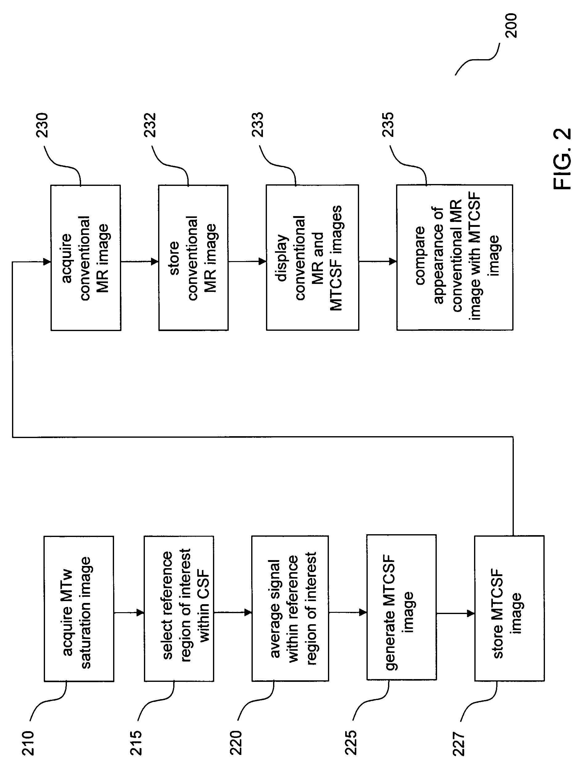
[0035]According to the present invention, an internally-referenced MTw image is generated, which is hereinafter referred to as an MTCSF image. An MTCSF image is generated on a voxel-by-voxel basis from a single acquired MTw image according to the following relation:

[0036]MTCSF=Mz⁡(ω)〈C⁢⁢S⁢⁢F〉ROI(2)

where MTCSF is the magnetization transfer ratio using CSF as the internal intensity reference; Mz(ω) is the MR signal acquired at the RF saturation frequency, ω, wherein Mz corresponds to the signal intensity in a given voxel within the single image; ROI is the MR signal intensity averaged over a region of interest (ROI) corresponding to cerebrospinal fluid (CSF) and normalized per voxel. Cerebrospinal fluid serves as an effective reference because it has negligible magnetization transfer effect, which is common across subjects, and is temporally invariant. Accordingly, instead of performing a separate reference scan and registering the saturation image to such a reference image, a quantit...


PUM


Abstract

Disclosed is a system and method for generating quantitative imagery of demyelination in the spinal cord. The method includes acquiring a magnetization transfer weighted (MTw) MR image of the spinal column, identifying a reference region of interest within the image corresponding to cerebrospinal fluid (CSF), averaging the signal intensity corresponding to the reference region of interest, and computing a ratio, on a voxel-by-voxel basis, of the signal intensity of each voxel by the averaged reference signal intensity. In doing so, normalized MTw images are obtained such that detrimental artifacts such as motion-induced errors, coil loading, and RF coil sensitivity variations are obviated.

Description

REFERENCE WITHIN THE IMAGE[0001]This application claims the benefit of U.S. Provisional Patent Application No. 60 / 602,158, filed on Aug. 17, 2004, which is hereby incorporated by reference for all purposes as if fully set forth herein.[0002]Research and development related to the invention disclosed below was funded in part through a grant from the National Institutes of Health / National Institute for Biomedical Imaging and Bioengineering, grant no. EB00991-01.BACKGROUND OF THE INVENTION[0003]1. Field of the Invention[0004]The present invention involves the field of Magnetic Resonance Imaging (MRI). Particularly, the present invention involves the use of MR signal information from a reference substance identified within an MR image to provide quantitative Magnetization Transfer weighted (MTw) images. More particularly, the present invention involves the use of MR signal corresponding to cerebrospinal fluid (CSF) as a reference to provide quantitative MTw images of the spinal cord.[00...

Claims


Application Information

Patent Timeline
Patent Type & Authority Patents(United States)
IPC IPC(8): G06K9/00
CPCA61B5/055G01R33/5605G06T7/0014G01R33/54G01R33/56509G01R33/5659G06T2207/10088G06T2207/20104G06T2207/30004A61B5/407
Inventor SMITH, SETHGOLAY, XAVIERVAN ZIJL, PETER C. M.
Owner THE JOHN HOPKINS UNIV SCHOOL OF MEDICINE
Why Eureka
Try Eureka