Ultrasound image enhancement and speckle mitigation method

a technology of ultrasound image and enhancement method, applied in the field of ultrasound imaging data processing techniques, can solve the problems of unsatisfactory enhancement effect, too much discontinuity at the edges of segmentation template, and interference with the doctor's diagnosis to some extent, so as to enhance ultrasound image, enhance ultrasound image, enhance ultrasound image

Active Publication Date: 2010-05-18
SHENZHEN MINDRAY BIO MEDICAL ELECTRONICS CO LTD
View PDF21 Cites 22 Cited by
  • Summary
  • Abstract
  • Description
  • Claims
  • Application Information

AI Technical Summary

Benefits of technology

[0007]According to another object of present invention, there's provided a new method of processing the feature region, which can make the enhanced image not susceptive of the image segmentation and avoid dependence of the enhancement effect on the segmentation template, so as not to produce an evident artificial boundary between the feature region and the non-feature region but to highlight some special information in the image and to remove or mitigate invalid information.
[0008]According to another object of present invention, there's provided a method of mitigating speckle noises, in particular black speckles in the ultrasound image.
[0013]C) merging the processed feature region and non-feature region to produce an enhanced image.
[0037]With the above technical solutions, the feature information in the ultrasonic image may be extracted and enhanced more effectively, and the image enhancement effect relies slightly upon the image segmentation, while it is advantageous to mitigate black speckles. The enhanced image is thus more suitable for the visual system of the human beings and more helpful for reference in medical diagnosis. The above and other advantages of present invention will be described with reference to the accompanying drawings.

Problems solved by technology

The speckle noises will mask some useful information in the image, thus interfering with the doctor's diagnosis to some extent.
The above prior art, however, has the following disadvantages: (1) although the method in the patent advantageously segments an image into a structural and a non-structural region, but it leaves out some inconspicuous features by only taking gradient scale information into account, in particular, the enhancement effect relies too much on the segmentation template when the structural region takes the gray-scale of the image as the sharpening coefficient, and too much discontinuity will occur at the edges of the segmentation template.
Accordingly the enhancement effect is not ideal; and (2) linear smoothing of the non-structural region cannot mitigate speckle noises greatly.

Method used


Image

  • Ultrasound image enhancement and speckle mitigation method
  • Ultrasound image enhancement and speckle mitigation method
  • Ultrasound image enhancement and speckle mitigation method

Examples


Embodiment Construction

[0055]Detailed descriptions will be made below to the invention, in conjunction with a preferred embodiment as shown in the accompanying drawings.

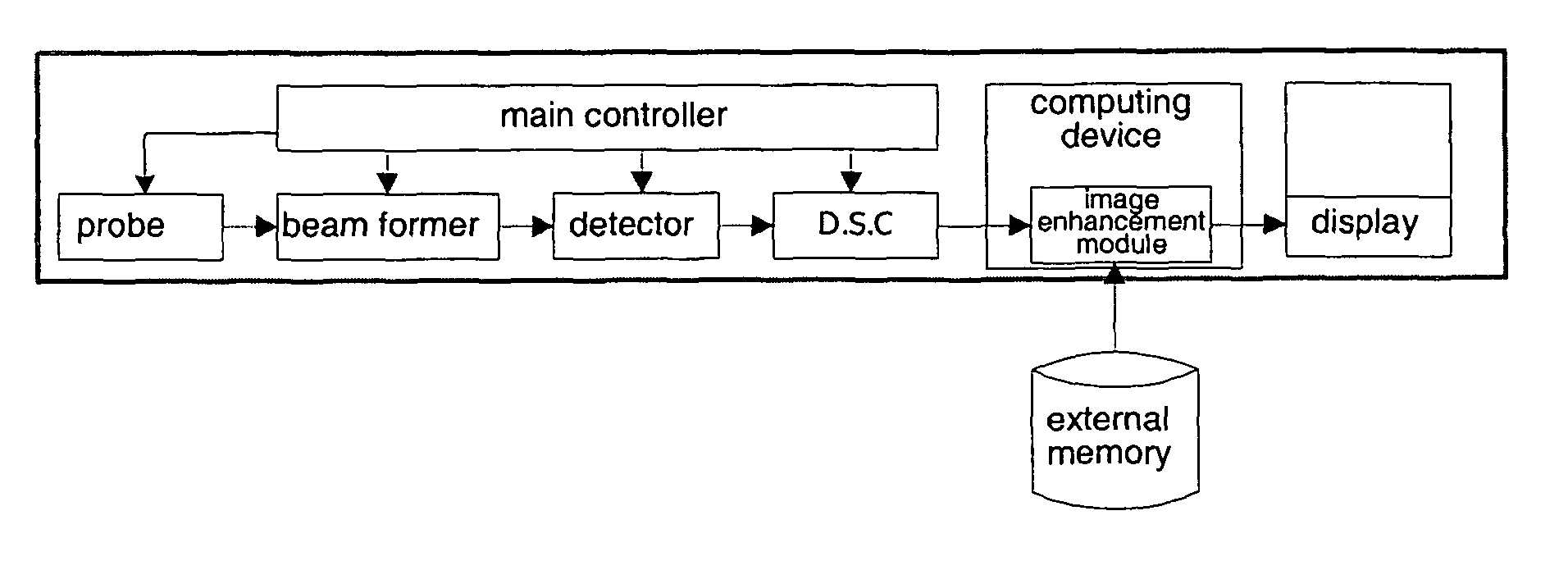
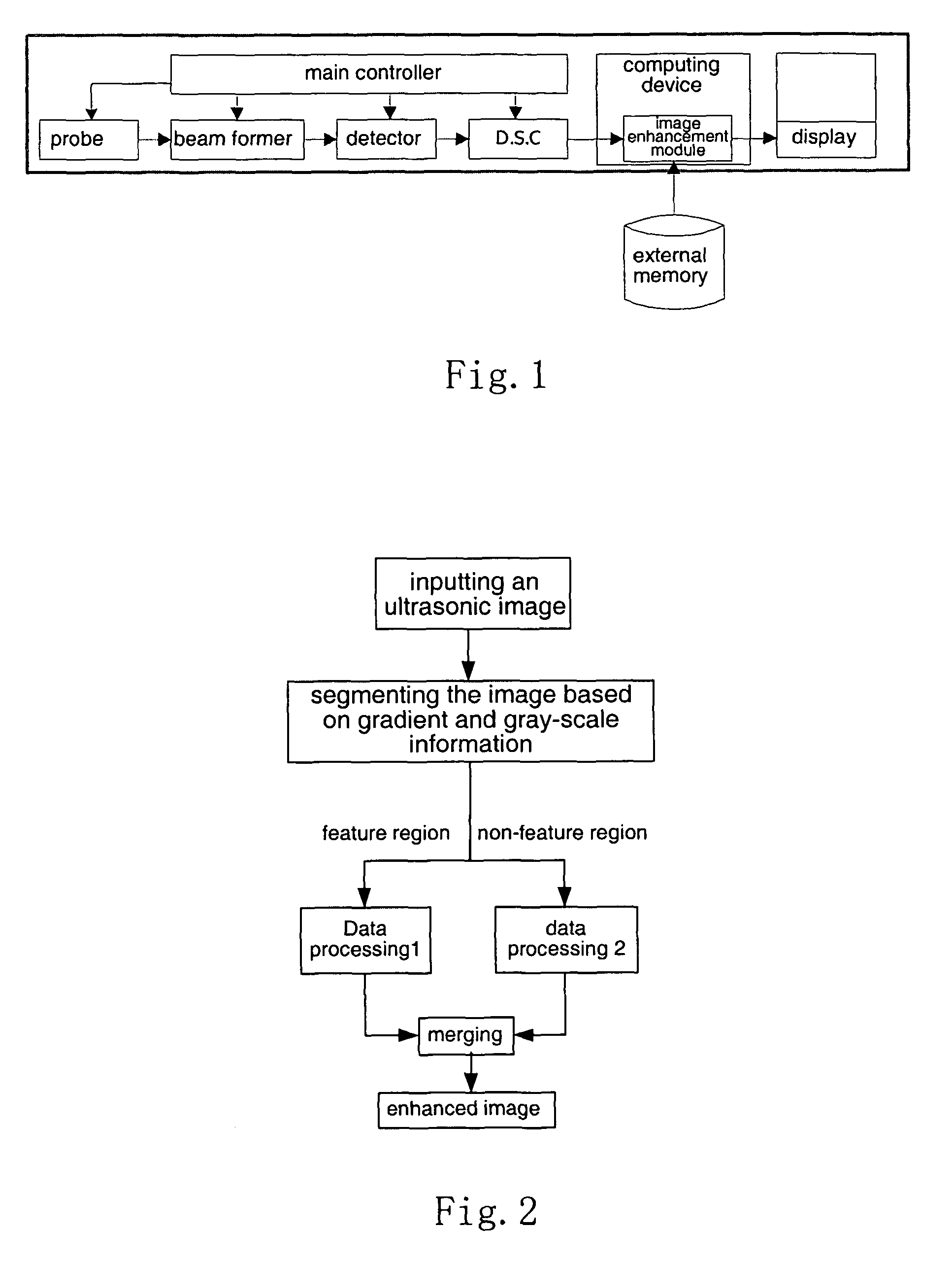
[0056]The invention can be implemented with the ultrasound imaging system shown in FIG. 1.

[0057]The method for enhancing an ultrasound image as provided in the invention is used by an ultrasound imaging system to optimize display of an ultrasound scanned image. As shown in FIG. 2, the system first reads the input ultrasound image data and then segments the image into a feature region and a non-feature region according to the gradient information and gray information in the image; sequentially, performing data processing (1) on the image data classified as the feature region and data processing (2) on the image data classified as the non-feature region, respectively. At last, the processed feature region and non-feature region are merged, to produce enhanced image data corresponding to the original ultrasound image, and then the enhanced im...


PUM


Abstract

A method for enhancing an ultrasound image is provided, wherein the ultrasound image is segmented into a feature region and a non-feature region, while sufficiently utilizing features contained in the ultrasound image, in particular including some inconspicuous features. The enhanced image according to present invention is not susceptive of the image segmentation and avoid dependence of the enhancement effect on the segmentation template, so as not to produce an evident artificial boundary between the feature region and the non-feature region but to highlight some special information in the image and to remove or mitigate invalid information. Thus the enhanced ultrasound image is particularly suitable for the visual system of the human beings.

Description

FIELD OF THE INVENTION[0001]The present invention relates to ultrasound techniques, and more particularly, to data processing techniques in ultrasound imaging, especially to an image enhancement and speckle mitigation method for improving the quality of ultrasound images.BACKGROUND OF THE INVENTION[0002]FIG. 1 shows a block diagram of a typical ultrasound imaging system. It operates as follows: under the control of a main controller, a probe transmits an ultrasound to a body tissue to be tested, and receives an echo signal reflected from the body tissue after a certain delay. The echo signal is fed into a beam former, which performs focus for delay, weighting and channel accumulation to generate signals on one or more scan lines. A detector detects the scanning signals output from the beam former and feeds them into a DSC (Digital Scanning Converter), where a coordinate conversion is implemented. The resulted image data is sent to a computing device (generally including a built-in p...

Claims


Application Information

Patent Timeline
Patent Type & Authority Patents(United States)
IPC IPC(8): G06K9/00
CPCG01S7/52034G01S7/52046G01S7/52063G01S7/52069G06T7/0083G06T7/0091G06T5/30G06T5/002G06T5/003G06T2207/10132G06T2207/20192G06T2207/30004G06T2207/20144G06T7/12G06T7/155G06T7/194
Inventor NI, DONGZHU, LEIHU, QINJUNFENG, HELIN
Owner SHENZHEN MINDRAY BIO MEDICAL ELECTRONICS CO LTD
Why Eureka
Try Eureka