Dose evaluating method and X-ray CT apparatus

a technology of x-ray ct and evaluating method, which is applied in the field of dose evaluating method and x-ray ct apparatus, can solve the problems of inability to evaluate whether the dose at the photography of an already reconstructed image is in excess, and the preliminary imaging becomes complex

Inactive Publication Date: 2010-07-13
GE MEDICAL SYST GLOBAL TECH CO LLC
View PDF25 Cites 8 Cited by
  • Summary
  • Abstract
  • Description
  • Claims
  • Application Information

AI Technical Summary

Benefits of technology

[0005]The conventional X-ray apparatus was capable of preventing a dose at the real imaging from becoming excessive for the subject.
[0014]In the dose evaluating method according to the third aspect, the operator has designated the dose less than or equal to the exhibited maximum dose. Thus, the dose can be prevented from becoming excessive for the subject.
[0021]In the dose evaluating method according to the sixth aspect, the operator has designated the dose less than or equal to the exhibited maximum dose. Thus, it is possible to prevent the dose from becoming excessive for the subject.

Problems solved by technology

However, there is a need to collect, by preliminary imaging, projection data corresponding to all views, which are necessary to reconstruct the preliminary image, thus causing a problem that the preliminary imaging becomes complex.
Also a problem arises in that it is not possible to evaluate whether a dose at the photography of an already reconstructed image is in excess.

Method used


Image

  • Dose evaluating method and X-ray CT apparatus
  • Dose evaluating method and X-ray CT apparatus
  • Dose evaluating method and X-ray CT apparatus

Examples


embodiment 1

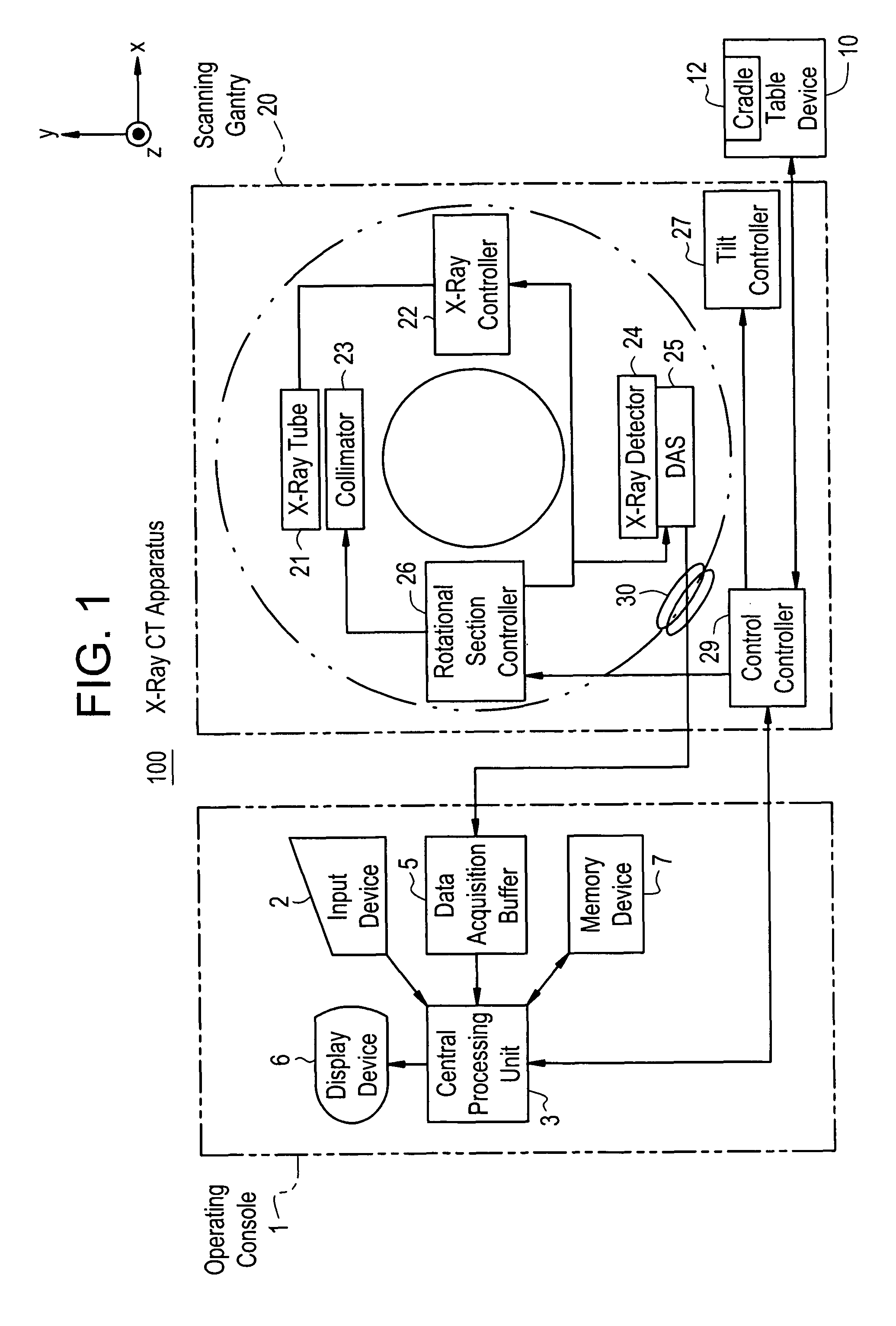
[0061]FIG. 1 is a schematic block diagram showing an X-ray CT apparatus according to an embodiment 1.

[0062]The X-ray CT apparatus 100 is equipped with an operating console 1, a table device 10 and a scanning gantry 20.

[0063]The operating console 1 is provided with an input device 2 which receives an operator's input, a central processing unit 3 which executes various processes, a data acquisition buffer 5 which collects projection data obtained by the scanning gantry 20, a display device 6 which displays a CT image or the like image-reconstructed from the projection data, and a memory device 7 which stores programs, data and an image therein.

[0064]The table device 10 includes a cradle 12 which places a subject thereon and inserts and draws it into and from a bore (cavity section). The cradle 12 is elevated and linearly moved by a motor built in the table device 10.

[0065]The scanning gantry 20 is provided with an X-ray tube 21, an X-ray controller 22, a collimator 23, an X-ray detect...

embodiment 2

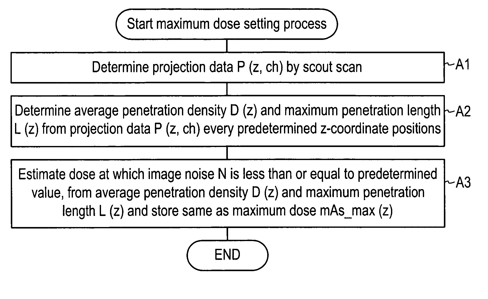
[0092]If the minimum value of the maximum dose mAs_max(z) at a linearly-moved range (range at each z-coordinate position) is assumed to be of a maximum dose where a helical scan is performed under a constant dose, then no dose is made excessive even at any point of the linearly-moved range. On the other hand, if the maximum value of the maximum dose mAs_max(z) at the linearly-moved range is assumed to be of a maximum dose, then image noise reaches less than equal to a predetermined value even at any point of the linearly-moved range.


PUM


Abstract

With the objective of evaluating whether a dose is excessive for a subject, such a dose that image noise reaches less than or equal to a predetermined value is estimated upon an axial scan on the basis of projection data acquired by a scout scan. The dose is set as a maximum dose. An area other than air, of an image obtained by the axial scan is partitioned into a plurality of small areas i. Image noises N(i) at the respective small areas i are calculated. A warning image G1 in which all of pixel values of pixels in small areas in which the image noises N(i) are less than or equal to a predetermined value, are substituted with pixel values expressed in black level, is created and displayed.

Description

CROSS REFERENCE TO RELATED APPLICATIONS[0001]This application claims the benefit of Japanese Application No. 2004-348730 filed Dec. 1, 2004.[0002]This application claims the benefit of Japanese Application No. 2004-348730 filed Dec. 1, 2004, incorporated by reference herein in its entirety.[0003]There has heretofore been known an X-ray apparatus which controls the output of an X-ray tube at the time of real imaging of a subject on the basis of a histogram indicative of pixel values for a preliminary image obtained by preliminarily imaging the subject by the X-ray apparatus (refer to, for example, a patent document 1).[0004][Patent Document 1] Japanese Patent Publication No. Hei 10(1998)-308899[0005]The conventional X-ray apparatus was capable of preventing a dose at the real imaging from becoming excessive for the subject.[0006]However, there is a need to collect, by preliminary imaging, projection data corresponding to all views, which are necessary to reconstruct the preliminary i...

Claims


Application Information

Patent Timeline
Patent Type & Authority Patents(United States)
IPC IPC(8): A61B6/00
CPCA61B6/032A61B6/488A61B6/542G01T1/02A61B6/03
Inventor GOHNO, MAKOTO
Owner GE MEDICAL SYST GLOBAL TECH CO LLC
Why Eureka
Try Eureka