

Lung cancer diagnosis using magnetic resonance imaging data obtained at three time points
a magnetic resonance imaging and cancer technology, applied in the field of medical imaging devices, can solve the problems of not increasing the overall survival rate in recent years, the overall survival rate of five years is only 14%, and the differentiation of benign from malignant tissue remains a challenge for the radiologist, etc., and achieves the effect of satisfying the imag
- Summary
- Abstract
- Description
- Claims
- Application Information
AI Technical Summary
Benefits of technology
Problems solved by technology
Method used
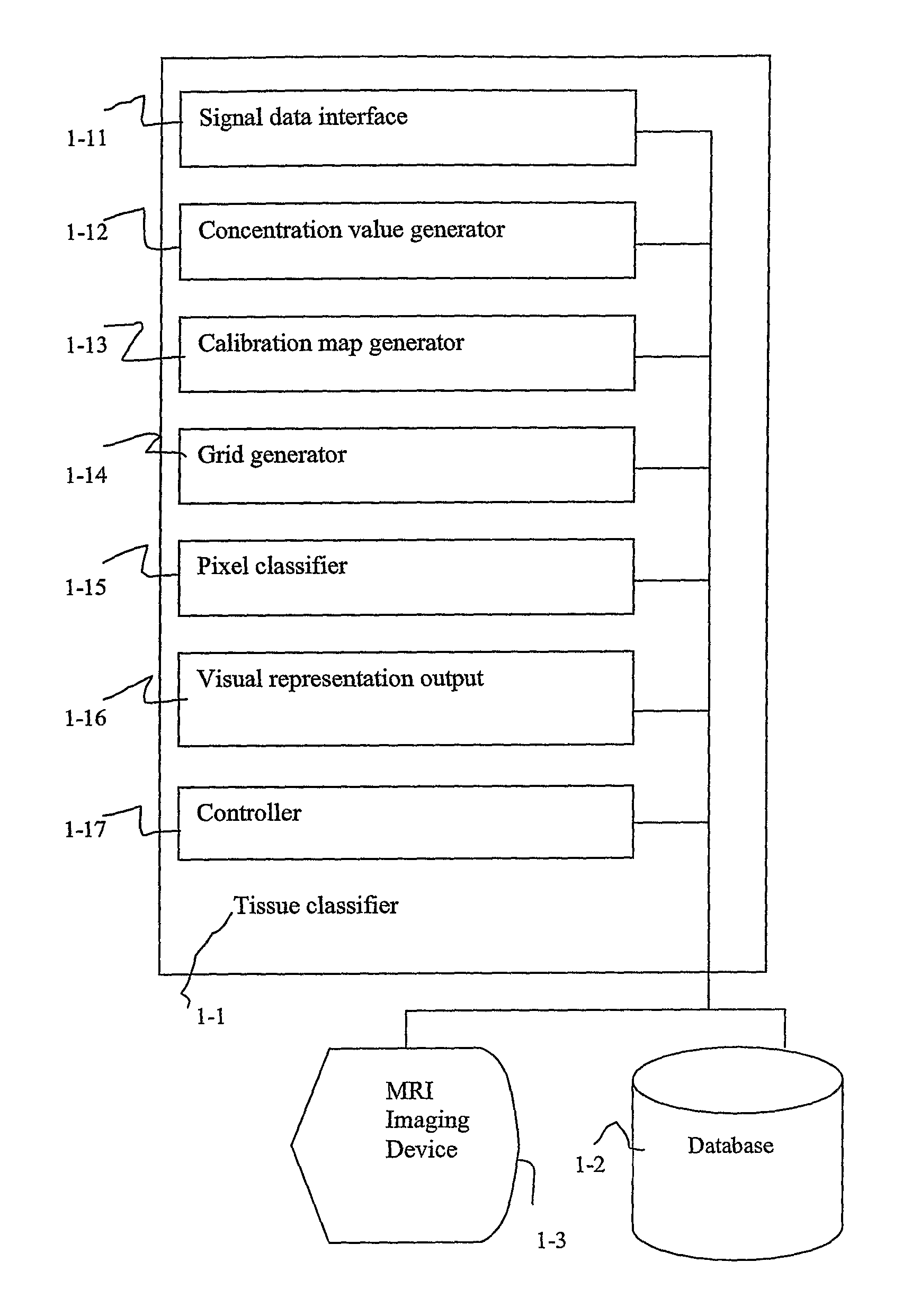
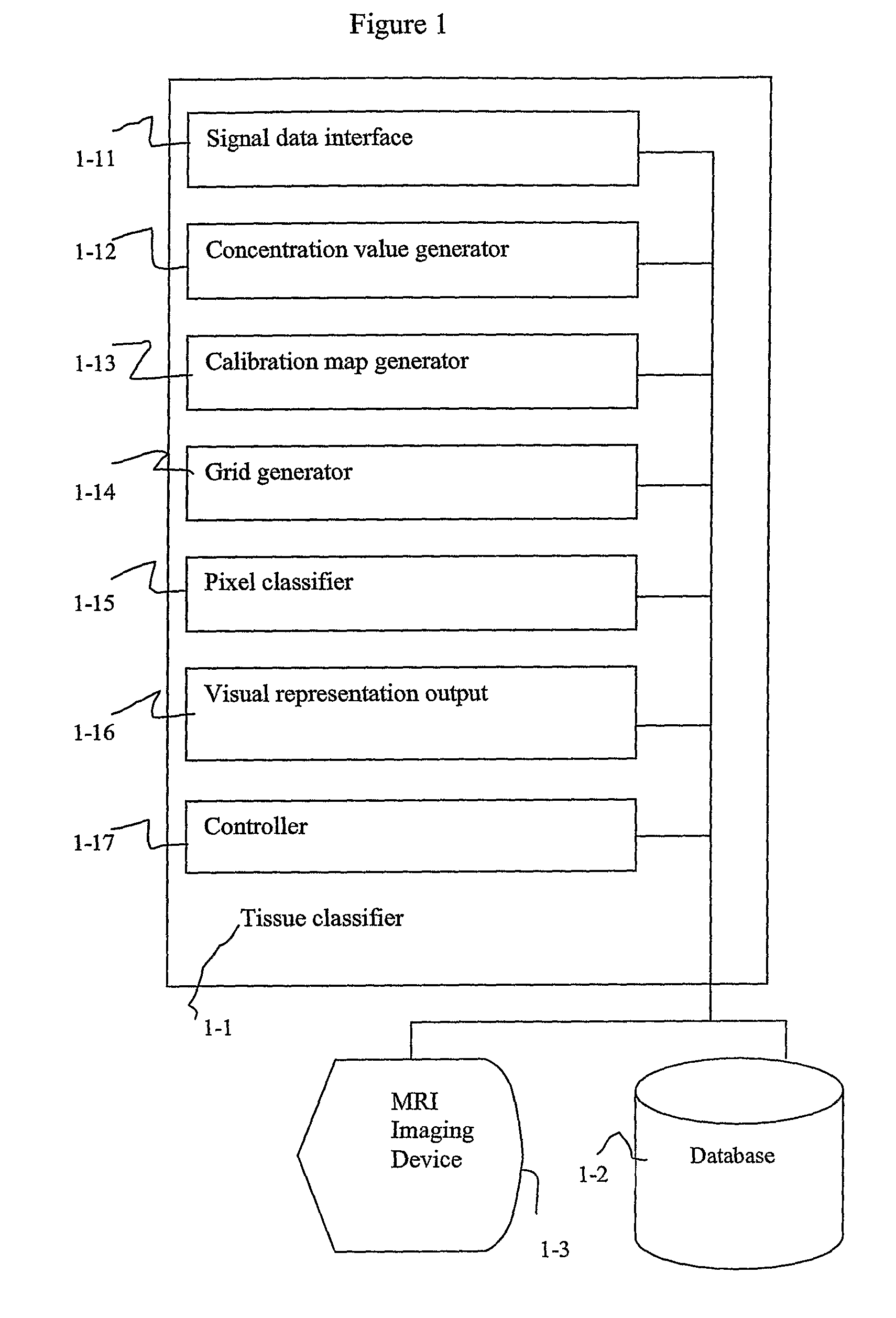
Image

Examples
Embodiment Construction
[0029]The following discussion and the foregoing figures describe embodiments of Applicant's invention as best understood presently by the inventor however, it will be appreciated that numerous modifications of the invention are possible and that the invention may be embodied in other forms and practiced in other ways without departing from the spirit of the invention. Further, features of embodiments described may be omitted, combined selectively, or as a whole, with other embodiments, or used to replace features of other embodiments, or parts thereof, without departing from the spirit of the invention. The figures and the detailed description are therefore to be considered as an illustrative explanation of aspects of the invention, but should not be construed to limit the scope of the invention.
[0030]Some of the conclusions stated in the following discussion are based on a study performed by the inventors in which dynamic contrast enhanced MRI experiments involved an animal model ...
PUM
 Abstract
Description
Claims
Application Information