Method of detection of platelet-activating antibodies that cause heparin-induced thrombocytopenia/thrombosis
a technology of platelet activation and thrombosis, which is applied in the field of detection of platelet activation antibodies that cause heparin-induced thrombocytopenia/thrombosis, to achieve the effects of reducing baseline platelet activation, increasing mfi, and confirming specificity of positive reactions
- Summary
- Abstract
- Description
- Claims
- Application Information
AI Technical Summary
Problems solved by technology
Method used
Image
Examples
example 1
and Clinical Testing
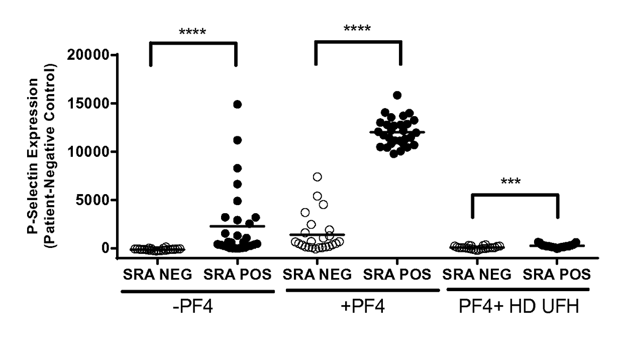
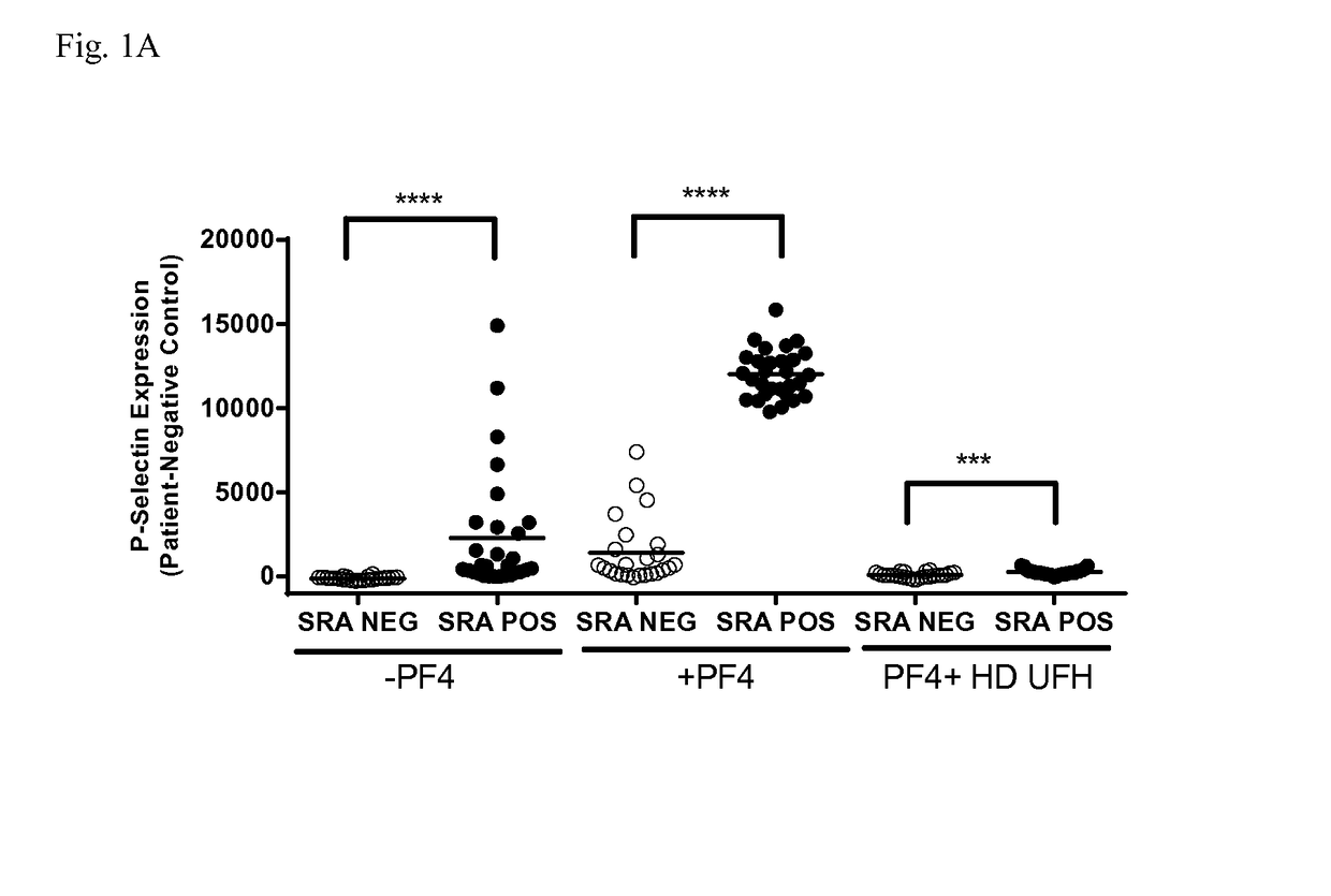
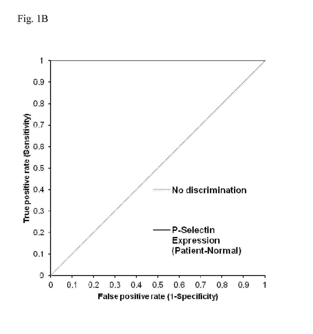
[0068]Serum samples (n=53) from patients suspected of having HIT were obtained from the Platelet and Neutrophil Immunology Laboratory of the BloodCenter of Wisconsin (BCW). Samples selected were PF4-ELISA positive with an optical density (OD) >0.4, that decreased ≧49% with high dose (HD; 100 U / ml) unfractionated heparin (UFH). SRA was performed at least twice on each sample and twenty-nine samples were consistently positive in the serotonin release assay (SRA) and 24 were negative. The research protocol was approved by the Institutional Review Board of BloodCenter of Wisconsin.
example 2
n Assay
[0069]Platelets were isolated from citrated platelet-rich plasmas from 2-3 Group O blood donors (to minimize inter-donor platelet variability) in the presence of prostaglandin E1. Reaction mixtures consisted of 1×10⁶ platelets in phosphate-buffered saline (PBS)-1% bovine serum albumin (BSA) to which was added (1) 50 μg / ml PF4, (2) 50 μg / ml PF4 and high dose (HD) UFH, or (3) buffer for 20 minutes at room temperature. The amount of PF4 added was optimized to maximize platelet activation in the assay. Ten microliters of serum was then added to produce a final reaction volume of 50 μl. Human PF4 was purified as previously described.
[0070]After incubation for 60 minutes at room temperature, phycoerythrin (PE)-labeled p-selectin antibody (BD Biosciences, San Jose, Calif.), and Alexa Fluor 647-labeled anti-GPIIb antibody 290.5 were added. After 15 minutes, the mixture was diluted in 200 μl of PBS-1% BSA and platelet events were acquired in an Accuri C6 flow cytometer. Events were ga...
example 3
bulin-Platelet Binding Assay
[0071]Platelets were isolated from citrated platelet-rich plasmas from 2-3 Group O blood donors (to minimize inter-donor platelet variability) in the presence of prostaglandin E1. Reaction mixtures consisted of 1×10⁶ platelets in phosphate-buffered saline (PBS)-1% bovine serum albumin (BSA) to which was added (1) 50 μg / ml PF4, (2) 50 μg / ml PF4 and high dose (HD) UFH, and (3) buffer for 20 minutes at room temperature. The amount of PF4 added was optimized to maximize IgG binding to platelets in the assay. 2.5 microliters of serum was then added to a final reaction volume of 50 μl. After incubation for 60 minutes at room temperature, FITC-labeled goat anti-human IgG antibody (Jackson Immunoresearch, West Grove, Pa.), and Alexa Fluor 647-labeled anti-GPIIb antibody 290.5 were added. After 30 minutes, the mixture was diluted in 200 μl of PBS-1% BSA and platelet events were acquired in an Accuri C6 flow cytometer. Events were gated by GPIIb positivity and anti...
PUM
| Property | Measurement | Unit |
|---|---|---|
| average molecular weight | aaaaa | aaaaa |
| temperature | aaaaa | aaaaa |
| volume | aaaaa | aaaaa |
Abstract
Description
Claims
Application Information
 - R&D
- Intellectual Property
- Life Sciences
- Materials
- Tech Scout
- Unparalleled Data Quality
- Higher Quality Content
- 60% Fewer Hallucinations
Browse by: Latest US Patents, China's latest patents, Technical Efficacy Thesaurus, Application Domain, Technology Topic, Popular Technical Reports.
© 2025 PatSnap. All rights reserved.Legal|Privacy policy|Modern Slavery Act Transparency Statement|Sitemap|About US| Contact US: help@patsnap.com


