Ultrasound diagnosis apparatus, image processing apparatus, image processing method, and image display method

A diagnostic device, ultrasonic technology, applied in the direction of acoustic wave diagnosis, ultrasonic/sonic wave/infrasonic wave diagnosis, infrasonic wave diagnosis, etc., which can solve the problems of increased workload for physicians, prolonged examination time, and deterioration of examination efficiency, etc.

Active Publication Date: 2013-03-06
TOSHIBA MEDICAL SYST CORP
View PDF2 Cites 0 Cited by
  • Summary
  • Abstract
  • Description
  • Claims
  • Application Information

AI Technical Summary

Problems solved by technology

[0014] However, in the above-mentioned "reinjection method", although it is possible to accurately differentiate liver tumors and diagnose the degree of malignancy, there is a problem that the physical burden on the subject and the workload on the physician increase due to the second administration of the ultrasound contrast medium. , and further, due to the extension of inspection time, the problem of deterioration of inspection efficiency

Method used


Image

  • Ultrasound diagnosis apparatus, image processing apparatus, image processing method, and image display method
  • Ultrasound diagnosis apparatus, image processing apparatus, image processing method, and image display method
  • Ultrasound diagnosis apparatus, image processing apparatus, image processing method, and image display method

Examples


Embodiment Construction

[0040] Preferred embodiments of the ultrasonic diagnostic apparatus, image processing apparatus, image processing method, and image display method related to the present invention will be described in detail below with reference to the drawings. In the following, an ultrasonic diagnostic apparatus incorporating an image processing device that executes the image processing method and the image display method related to the present invention will be described as an example.

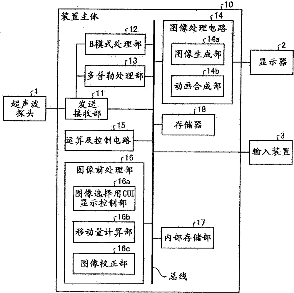
[0041] First, use figure 1 , the configuration of the ultrasonic diagnostic apparatus in Embodiment 1 will be described. figure 1 It is a diagram for explaining the configuration of the ultrasonic diagnostic apparatus in Example 1. Such as figure 1 As shown, the ultrasonic diagnostic apparatus in this embodiment has an ultrasonic probe 1 , a monitor 2 , an input device 3 , and an apparatus main body 10 .

[0042] The ultrasonic probe 1 incorporates an ultrasonic vibrator that gathers a plurality of vi...


PUM


Abstract

When an image creating unit creates a three-dimensional ultrasound image of a blood-vessel early phase, an operator presses an image selecting button, and selects an area to be displayed as superimposed by using an image-selecting Graphical User Interface (GUI) displayed by an image-selecting GUI display-control unit. After the image creating unit start creation of a three-dimensional ultrasound image in a late phase, when the operator presses a composition display button, the movement-distance calculating unit calculates a movement distance between a three-dimensional tissue image at the moment of the press of the composition display button and each of three-dimensional tissue images in the area to be displayed as superimposed, and an image correcting unit corrects each of three-dimensional contrast enhanced images corresponding the three-dimensional tissue images of the blood-vessel early phase by using the movement distance. An animated-image compositing unit creates a group of images for animation that the corrected three-dimensional contrast enhanced images of the blood-vessel early phase are each combined with a three-dimensional contrast enhanced image of the late phase, and a monitor displays the group of the images for animation in animation.

Description

technical field [0001] This application enjoys the benefit of priority of Japanese Patent Application No. 2009-137703 filed on June 8, 2009 and Japanese Patent Application No. 2010-108582 filed on May 10, 2010, which are incorporated herein by reference the entire content of . [0002] The present invention relates to an ultrasonic diagnostic device, an image processing device, an image processing method, and an image display method. Background technique [0003] Conventionally, in ultrasonic examinations using ultrasonic diagnostic equipment, contrast-enhanced ultrasonographic examinations ( For example, refer to "Medical Imaging and Radiation Equipment Handbook" edited by Japan Imaging Medical System Industry Association, Nago Art Printing Co., Ltd., 2013, p.221-225). [0004] Here, it is recommended that in abdominal contrast ultrasonography for the purpose of differentially diagnosing "benign and malignant" liver tumors, after administering an intravenously injectable ...

Claims


Application Information

Patent Timeline
Patent Type & Authority Patents(China)
IPC IPC(8): A61B8/06
CPCA61B8/488A61B8/08A61B8/5253A61B8/085A61B8/4254G01S7/52074A61B8/06A61B8/14A61B8/481A61B8/4209A61B8/13G06T7/2013A61B8/483A61B8/0891A61B8/4218G06T7/223
Inventor 小笠原洋一西野正敏小林丰郡司隆之
Owner TOSHIBA MEDICAL SYST CORP
Why Eureka
Try Eureka