Method and kit for examining complement system

a complement system and kit technology, applied in the field of methods and kits for examining the complement system, can solve the problems of variation in the sensitivity of sheep erythrocytes, excessive activation of the complement system, and imposing severe tissue damage on an organism

Inactive Publication Date: 2018-02-22
NAT UNIV ASAHIKAWA MEDICAL UNIV
View PDF0 Cites 1 Cited by
  • Summary
  • Abstract
  • Description
  • Claims
  • Application Information

AI Technical Summary

Benefits of technology

[0024]Since the method and the kit according to one or more embodiments of the present invention do not require serial dilution of a blood sample, and do not use hemolysis of ovine erythrocytes as an index, they do not have the problems of complicated operation and measurement errors caused thereby, and variation among rots. The method and the kit of the present invention can detect both compleme

Problems solved by technology

While the complement system is originally a biological defense mechanism against invasion of pathogens, excessive activation of the complement system can impose severe tissue damage on an organism because MAC also causes lysis of cells other than pathogens, for example own cells of the organism.
While CH50 has been widely used as a method for measuring a complement titer, it has the problems including complicated operation in serial dilution of the test sample, occurrence of a measurement error caused by the serial dilution, and variation in the sensit

Method used


Image

  • Method and kit for examining complement system
  • Method and kit for examining complement system
  • Method and kit for examining complement system

Examples


example 1

Evaluation of Binding Between CL-P1 and PTXs

1-1. Evaluation Using ELISA System

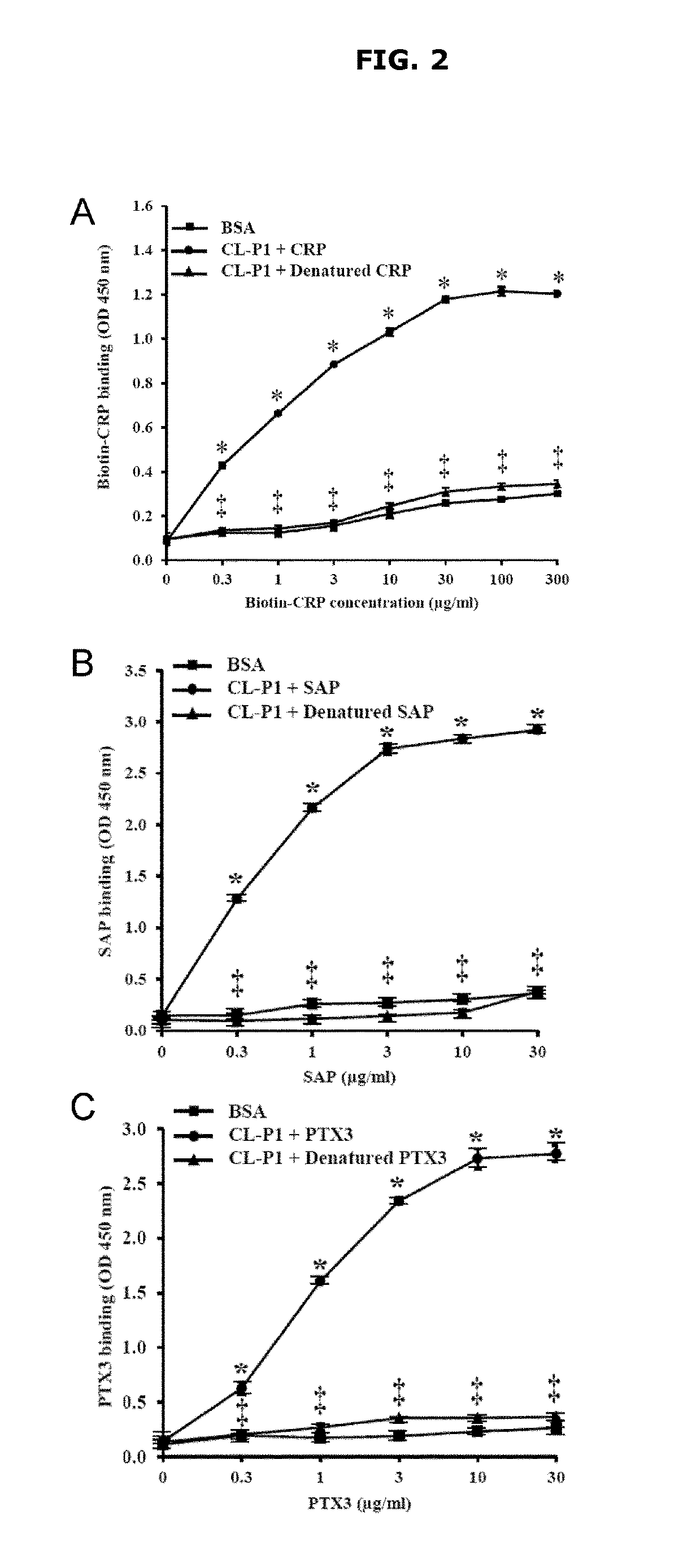
[0138]Binding between CL-P1 extracellular domain and PTXs (CRP, SAP and PTX3) was evaluated by using ELISA. The results are shown in FIGS. 2A to 2C. CRP, SAP and PTX3 bind to CL-P1 extracellular domain specifically and dose-dependently, and they lost the ability to bind to CL-P1 extracellular domain when they were heat-denatured.

1-2. Evaluation Using Cell System

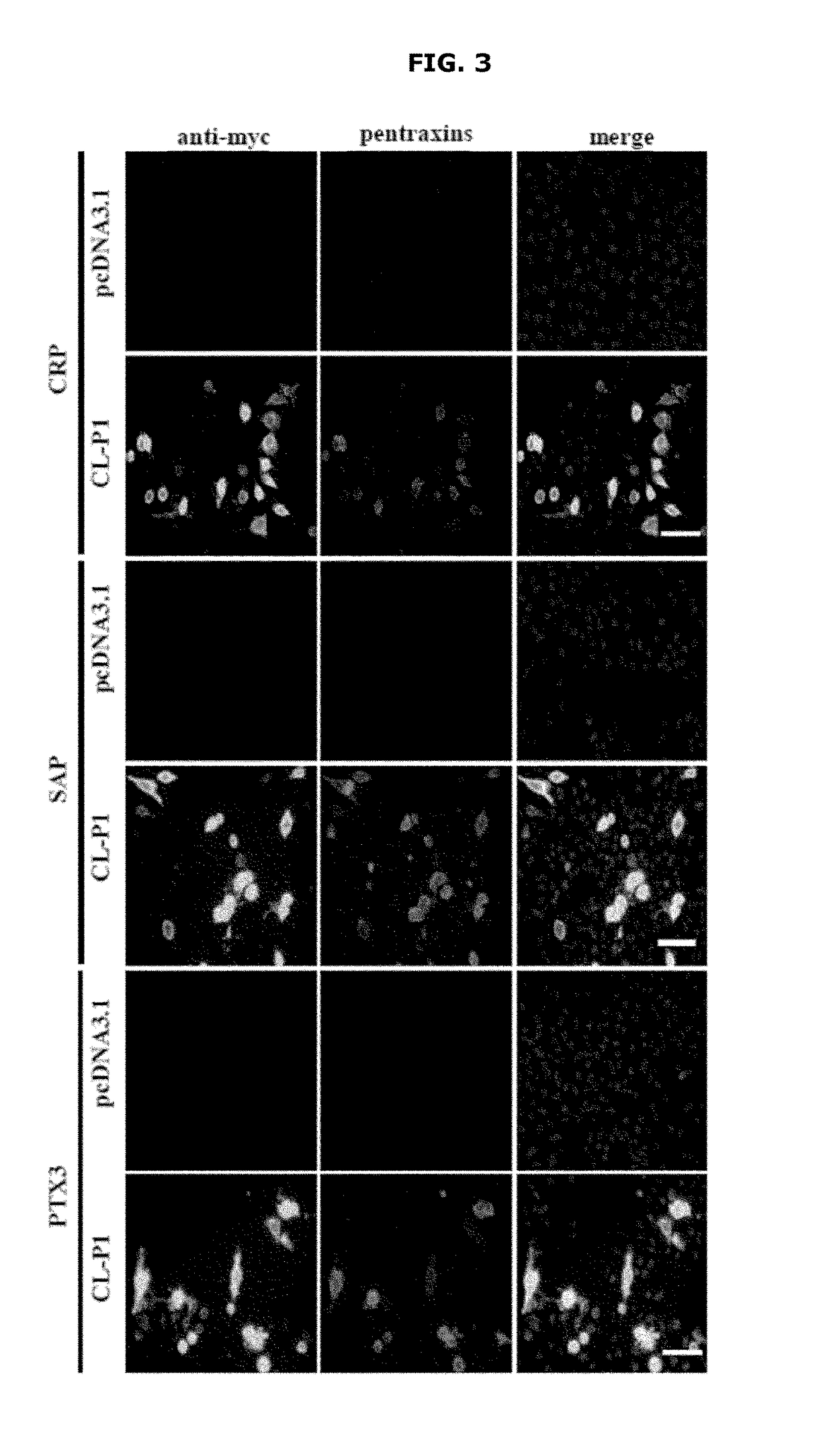
[0139]Binding between CL-P1 and PTXs was evaluated by using CHO / ldlA7 cells. As shown in FIG. 3, while CRP, SAP and PTX3 were co-localized with CL-P1 in CL-P1 expression cells, none of CRP, SAP, and PTX3 was detected in the cells expressing pcDNA3.1 control vector. This indicates that CRP, SAP and PTX3 bind to CL-P1 on the cell surface. A mean fluorescent intensity was calculated from each fluorescent image, and a binding amount of CRP, SAP or PTX3 relative to the CL-P1 expression amount was compared. The binding amount of CRP to CL-P1 was in a lowe...

example 2

Evaluation of Complement Activation Early-Phase Reaction Induced by Binding Between CL-P1 and PTXs

2-1. Evaluation of Complement Activation Early-Phase Reaction Using ELISA System

[0140]Induction of complement activation early-phase reaction by binding between CL-P1 extracellular domain and PTXs was evaluated by using ELISA. As shown in FIG. 5, in the presence of human complement serum, increase in C3d deposition amount depending on addition of CRP was observed in a CL-P1 extracellular domain-immobilized well. When C1q depleted serum was used, C3d deposition amount in a CL-P1 extracellular domain-immobilized well increased by addition of CRP, SAP or PTX3, and this increase in C3d deposition amount was further enhanced by using C1q depleted serum supplemented with C1q (FIG. 6). These indicate that CRP, SAP and PTX3 bind to extracellular domain of CL-P1, and activate the complement activation early-phase reaction via C1q to induce deposition of C3d.

2-2. Evaluation of Complement Activati...

example 3

Evaluation of Complement Activation Amplifying Reaction Induced by Binding Between CL-P1 and PTXs

3-1. Evaluation of Complement Activation Amplifying Reaction Via CFB Using ELISA System

[0142]Induction of the complement activation amplifying reaction by binding between CL-P1 extracellular domain and PTXs was evaluated by using ELISA. When CFB depleted serum supplemented with CFB was used, further increase in C3d deposition amount depending on the addition of CRP, SAP and PTX3 in a CL-P1 extracellular domain-immobilized well was observed, as compared with the case where CFB depleted serum was used (FIG. 11). These indicate that CRP, SAP and PTX3 bind to extracellular domain of CL-P1, and activate the complement activation amplifying reaction via CFB to induce deposition of C3d.

3-2. Evaluation of Complement Activation Amplifying Reaction Via CFB Using Cell System

[0143]Induction of complement activation amplifying reaction by binding between CL-P1 and PTXs was evaluated by using CL-P1 ex...


PUM


Abstract

A method for examining a complement system of a subject, including: a reaction step of letting a scavenger receptor or a functional equivalent thereof, a pentraxin family protein or a functional equivalent thereof and a blood sample collected from the subject coexist in vitro; and a detection step of detecting complement activation amplifying reaction and/or complement activation late-phase reaction induced by interaction between the scavenger receptor or the functional equivalent thereof and the pentraxin family protein or the functional equivalent thereof from a reactant after the reaction step. Also provided are a kit for examining a complement system, and a method for testing a test substance for the ability to activate or suppress a complement system.

Description

CROSS-REFERENCE TO RELATED APPLICATIONS[0001]This application claims priority to Japanese application No. 2016-159453, filed Aug. 16, 2016, the content of which is incorporated herein by reference in its entirety for all purposes.TECHNICAL FIELD[0002]The present invention relates to a method for examining activation of the complement system and / or abnormality of the complement system of a subject, and a kit for the examination.BACKGROUND OF THE INVENTION[0003]The complement system is a kind of innate immunity that is activated in response to invasion of a pathogen, and is composed of a large number of complement proteins that exist as membrane-bound proteins or soluble proteins in blood.[0004]The complement system activating pathways are roughly classified into three pathways: a classical pathway that is specifically activated by antigen-antibody reaction against a pathogen, a secondary pathway that is not accompanied by a mechanism of recognizing a pathogen, and a lectin pathway th...

Claims


Application Information

Patent Timeline
IPC IPC(8): G01N33/557G01N33/50
CPCG01N33/557G01N2333/4716G01N33/502G01N33/5008G01N33/6893
Inventor WAKAMIYA, NOBUTAKAOHTANI, KATSUKI
Owner NAT UNIV ASAHIKAWA MEDICAL UNIV
Why Eureka
Try Eureka