
Targeted magnetic resonance imaging (MRI) contrast agent and preparation method thereof

A contrast agent and targeting technology, applied in the field of biomedical materials, can solve the problems of target reduction, identification, phagocytosis and degradation, and retention of negative MRI contrast agents, and achieve the effect of avoiding immunogenic defects

Inactive Publication Date: 2014-04-02
海南医学院附属医院
View PDF1 Cites 0 Cited by
  • Summary
  • Abstract
  • Description
  • Claims
  • Application Information

AI Technical Summary

Problems solved by technology

However, the molecular weight of the antibody linked to the negative MRI contrast agent is usually relatively large, with strong immunogenicity, and is easily recognized, phagocytized and degraded by the human immune system, which greatly reduces the targeting of the negative MRI contrast agent. Contrast agents basically stay in the laboratory stage
In addition, different tumor cell membranes express different specific proteins, so the antibodies carried by negative MRI contrast agents can only be used for the diagnosis of one tumor, and the scope of application is relatively narrow

Method used


Image

  • Targeted magnetic resonance imaging (MRI) contrast agent and preparation method thereof
  • Targeted magnetic resonance imaging (MRI) contrast agent and preparation method thereof
  • Targeted magnetic resonance imaging (MRI) contrast agent and preparation method thereof

Examples


Embodiment 1

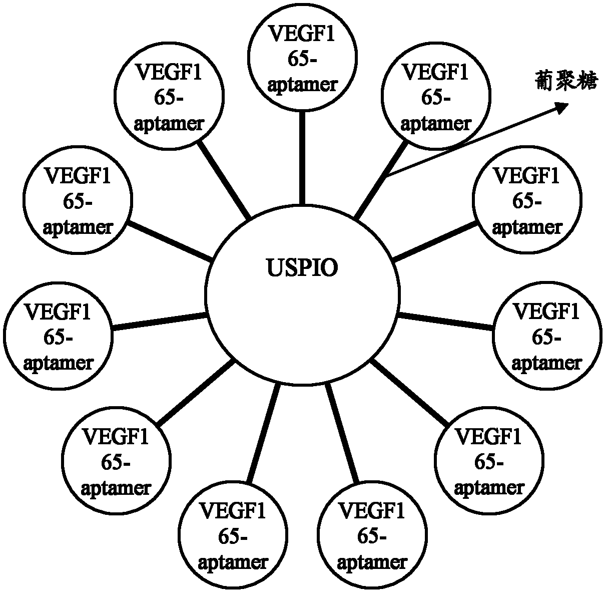
[0048] Embodiment 1: Preparation of MRI contrast agent of the present invention

[0049] Dissolve 10g of Dextran T-40 in 50ml of water, heat to 70°C, stir, and add 5ml of 10mol / L NaOH at the same time to react for 5h, then add 3ml of 5mol / L chloroacetic acid, keep the temperature at 70°C for 20min, and wait for the reaction system After cooling, adjust pH=8 with dilute hydrochloric acid; precipitate the reactant with methanol, purify in styrene-type anion-cation resin (the ratio of anion and cation resin is 1.5:1), and concentrate in vacuum at 50°C on a rotary evaporator , and then freeze-dry the product to obtain dextran T-40 with a carboxyl group, which is ready for use.

[0050] Prepare 5ml of 1mol / L FeCl 3 ·6H 2 O, and add 660mg FeCl 2 4H 2 O, N 2 Protected for use. Weigh 5.0 g of the above-mentioned dextran T-40 with carboxyl groups and dissolve it in 15 mL of once-distilled water, N 2 Heating to 75°C under protection, adding the prepared FeCl 3 ·6H 2 O-FeCl 2 4...

Embodiment 2

[0052] Embodiment 2: the coupled detection of MRI contrast agent of the present invention

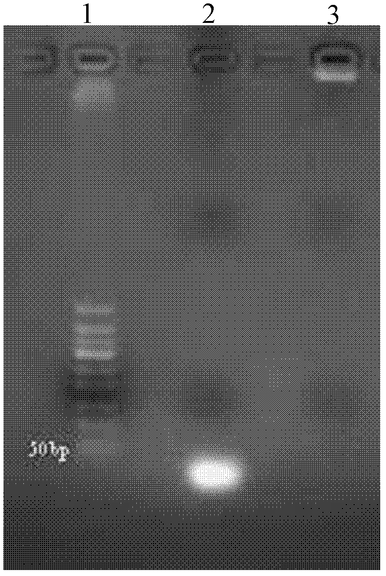
[0053] The MRI contrast agent prepared in Example 1 and the RNA of the present invention are carried out by 1.8% agarose gel electrophoresis, the results are shown in figure 2 . Depend on figure 2 It can be seen that the size of the RNA band that is not coupled to the superparamagnetic iron oxide particles coated with carboxydextran T-40 is below 50 bp, which is consistent with the actual situation. And the band after coupling hinders its migration in electrophoresis due to the cross-linked superparamagnetic iron oxide particles coated with carboxydextran T-40, and is close to the sample hole, thus indicating that the MRI of the present invention Contrast successful coupling.

Embodiment 3

[0054] Embodiment 3: In vitro targeted cell proliferation test of MRI contrast agent of the present invention

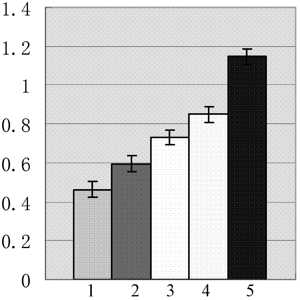
[0055] Experimental design: establish blank group, MRI contrast agent high-dose group of the present invention, MRI contrast agent middle-dose group of the present invention, MRI contrast agent low-dose group of the present invention and non-targeting negative MRI contrast agent (phenanthrene Limag) group.

[0056]The same amount of human umbilical vein endothelial cells (HUVEC cells) was added to 5 groups, VEGF165 was not added to the blank group, and the same amount of VEGF165 was added to the remaining 4 groups to stimulate the growth of HUVEC cells. 50 μL, 25 μL, and 12.2 μL were added to the high, medium, and low groups, respectively. The MRI contrast agent of the present invention, Feridex group adds 50 μ L Feridex, uses microplate reader statistical detection result, see image 3 .

[0057] Depend on image 3 It can be seen that the optical density of HUVEC...


PUM


Abstract

The invention relates to the field of biomedical materials, and discloses a targeted magnetic resonance imaging (MRI) contrast agent and a preparation method thereof. The method for preparing the MRI contrast agent comprises the following step of: covalently coupling superparamagnetic iron oxide particles of which the outer layers are wrapped by carboxyl glucosan and ribonucleic acid (RNA) which is modified by amino at the 5' terminal of a sequence shown as SEQ ID No:1, wherein the amino at the 5' terminal of the RNA is covalently coupled with carboxyl on glucosan. The MRI contrast agent is a negative contrast agent, the RNA of which the amino is introduced at the 5' end of the sequence shown as SEQ ID No:1 and which is connected to the contrast agent is modified human vascular endothelial cell growth factor 165 (VEGF165)-Aptamer, so that the VEGF165-Aptamer can be targeted to VEGF165 in tumor cells, overcome the defect of immunogenicity and can be applied to various kinds of tumor diagnosis.

Description

technical field [0001] The invention relates to the field of biomedical materials, in particular to a targeted MRI contrast agent and a preparation method thereof. Background technique [0002] MRI, that is, magnetic resonance imaging, is called Magnetic Resonance Imaging in English, also known as nuclear magnetic resonance imaging. MRI uses the principle of nuclear magnetic resonance, according to the different attenuation of the released energy in different structural environments inside the material, and through the detection of the emitted electromagnetic waves by an external gradient magnetic field, the position and type of the atomic nucleus that constitutes the object can be known. Drawn as a structural image of the interior of the object. [0003] Various tissues of the human body contain a large amount of water and hydrocarbons, so hydrogen nuclei are the first choice for human MRI due to their high flexibility and strong signal. The signal intensity of NMR is rel...

Claims


Application Information

Patent Timeline
Patent Type & Authority Patents(China)
IPC IPC(8): A61K49/10
Inventor 涂蓉尤晓光白玉杰
Owner 海南医学院附属医院
Learn More