PD-1 antibody detection kit and application thereof

A PD-1 and antibody detection technology, which is applied in the direction of measuring devices, instruments, scientific instruments, etc., can solve the problems that the specific detection of PD-1 antibodies cannot be realized, and achieve a wide range of applications, simple methods of use, and high specificity Effect

Inactive Publication Date: 2015-09-23
XIEHE HOSPITAL ATTACHED TO TONGJI MEDICAL COLLEGE HUAZHONG SCI & TECH UNIV
View PDF2 Cites 5 Cited by
  • Summary
  • Abstract
  • Description
  • Claims
  • Application Information

AI Technical Summary

Problems solved by technology

[0006] The invention provides a PD-1 antibody detection kit and its application, which are used to solve technical defects such as the inability to realize the specific detection of PD-1 antibody by the double-antibody sandwich method in the prior art

Method used


Image

  • PD-1 antibody detection kit and application thereof
  • PD-1 antibody detection kit and application thereof
  • PD-1 antibody detection kit and application thereof

Examples


Embodiment 1PD-1

[0037] Example 1PD-1 Antibody Detection Kit

[0038] 1. Materials and sources:

[0039] Antibody capture agent: a recombinant protein of the extramembrane domain of mouse PD-1 cells, produced by ACRO Biosystems, the article number is PD1-M5228, and its amino acid sequence is shown in SEQ ID NO.1;

[0040] Solid phase carrier: ELISA plate, produced by Coster;

[0041] Labeled antibody: HRP-coupled goat anti-rat IgG antibody, produced by KPL Company, the catalog number is 141612.

[0042] 2. PD-1 Antibody Detection Kit

[0043] The PD-1 antibody detection kit of this example consists of the above-mentioned solid phase carrier, antibody capture agent and labeled antibody, which are individually packaged and placed in a box.

Embodiment 2

[0044] Embodiment 2 PD-1 antibody detection kit and preparation method thereof

[0045] The PD-1 antibody detection kit in this example is an improvement based on the PD-1 antibody detection kit in Example 1, which pre-coats the antibody capture agent on a solid phase carrier to form a pre-coated solid phase. Phase carrier, the kit consists of the above pre-coated solid phase carrier, labeled antibody and the following components:

[0046] PD-1 antibody standard product: produced by American Bioxcell Company, the clone number is RMP1-14;

[0047] Coating buffer: Na per liter 2 CO 3 1.59g, NaHCO 3 2.93g, pH value is 9.6;

[0048] Wash buffer (PBS): 8.0g NaCl, 0.2g KCl, KH per liter 2 PO 4 0.24g, Na 2 HPO 4 12H 2 O 3.628g, the pH value is 7.4; Tween-200.5mL (PBST) can be added further;

[0049] Blocking solution: add 1 mg / mL bovine serum albumin (BSA, produced by Sigma) to the above PBS;

[0050] Chromogenic solution: produced by KPL Company, the article number is ...

Embodiment 3

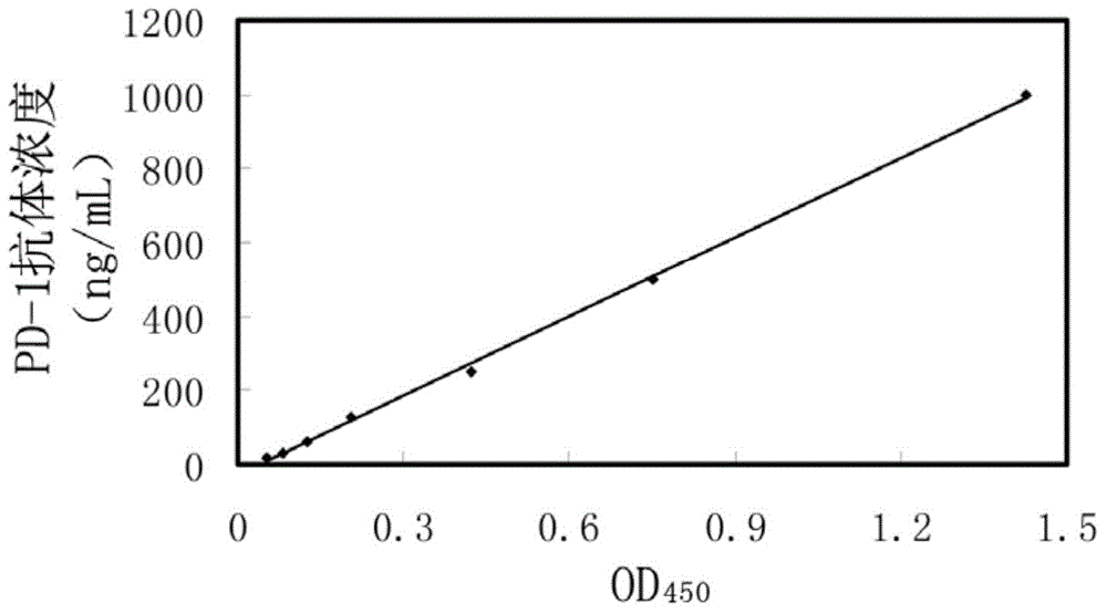
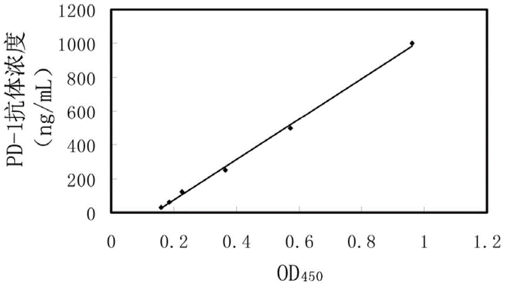
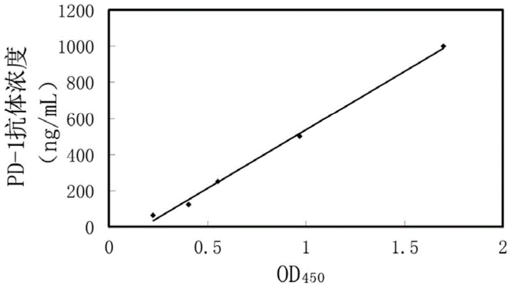
[0056] Example 3 Detection of PD-1 Antibody Concentration in Physiological Saline

[0057] The PD-1 antibody detection kit of Example 2 is used for detection, and the method is specifically as follows:

[0058] 1. Dilute the above PD-1 antibody standard with normal saline to concentrations of 1000ng / mL, 500ng / mL, 250ng / mL, 125ng / mL, 62.5ng / mL, 31.25ng / mL and 15.625ng / mL Gradient dilution; In addition, use the above PBST to add 0.2 μL of HRP-coupled goat anti-rat IgG antibody per mL to prepare the labeled antibody working solution.

[0059] 2. Take 100 μL of each gradient dilution solution, add them to the wells of the above-mentioned pre-coated ELISA plate, seal the plate, and incubate at 37°C for 1 hour.

[0060] 3. Pour off the solution in the ELISA plate, wash the plate 5 times with the above-mentioned washing buffer PBST, after clapping the plate, add 100 μL of the above-mentioned labeled antibody working solution to each well, seal the plate, and incubate at 37°C for 30 ...


PUM

PropertyMeasurementUnit
concentrationaaaaaaaaaa

Abstract

The invention provides a PD-1 antibody detection kit and application thereof. The PD-1 antibody detection kit comprises a solid phase carrier, an antibody trapping agent and a labeled antibody, wherein the antibody trapping agent is a recombinant protein of a PD-1 cytomembrane outer structure, and the amino acid sequence of the antibody trapping agent is SEQ ID NO.1. The PD-1 antibody detection kit can quickly detect a PD-1 antibody in a to-be-detected sample, such as in-vitro serum and tissues homogenate, the specificity of detection is high, the sensitivity of detection is high, and the application range is wide.

Description

technical field [0001] The invention relates to an antibody detection kit, in particular to a PD-1 antibody detection kit and its application. Background technique [0002] Under normal circumstances, specific cytotoxic T lymphocytes (CTL) generated by the activation of the body's immune response can kill tumor cells in the body. Tumor immunosuppression is an important mechanism for tumor cells to evade killing by the body's immune system. Recent studies in the field of tumor immunology have found that programmed death-1 (PD-1)-mediated immunosuppressive response is an important reason for CTL apoptosis, weakness, and functional exhaustion. Tumor cells and their tumor-associated immunosuppressive cells express B7-H1 / 4 molecules on the surface, which are known PD-1 ligands. When CTL enters the tumor microenvironment, PD-1 on the surface of CTL cells will be bound by it The activation of the body triggers the phosphorylation of SHP1 / 2 in the cell, which leads to apoptosis or...

Claims


Application Information

Patent Timeline
Patent Type & Authority Applications(China)
IPC IPC(8): G01N33/543
CPCG01N33/543
Inventor 王琳王征李永奎方敏张剑祁闪闪
Owner XIEHE HOSPITAL ATTACHED TO TONGJI MEDICAL COLLEGE HUAZHONG SCI & TECH UNIV
Why Eureka
Try Eureka