Electrical Impedance Tomography Lung Imaging Method Based on 3D Accelerometer

A technology of electrical impedance tomography and imaging method, applied in diagnosis, medical imaging, diagnostic recording/measurement, etc., can solve the problems of positive end-expiratory pressure, large error, long monitoring time, etc., and achieve high calculation accuracy. Effect

Active Publication Date: 2019-04-26
合肥诺和电子科技有限公司
View PDF4 Cites 0 Cited by
  • Summary
  • Abstract
  • Description
  • Claims
  • Application Information

AI Technical Summary

Problems solved by technology

One of the characteristics of these places is that the monitoring time is long and the patient's posture is uncertain; in addition, there is another characteristic of these places that the patient is mechanically ventilated, and this ventilation method will affect the positive end-expiratory pressure (PEEP)
The traditional impedance tomography (EIT) lung imaging system usually adjusts the patient's posture through manual input by the doctor. The doctor estimates the angle according to the current posture relative to the normal lying posture. At the same time, after each posture is determined, it needs to Re-modeling evaluation calculation, this method will mainly cause the following problems: 1. The input of the doctor's posture data is not very accurate, it is estimated by the doctor himself, and the error is large; 2. During the operation or in the ICU During the monitoring process, the patient may change posture frequently, and the posture change requires timely re-input by the doctor, which brings inconvenience to the use; 3. Traditional equipment needs to be re-modeled and calculated after each input of posture information to achieve a real Imaging, resulting in insufficient continuity in the monitoring process; 4. In the ICU room, because the cycle is very long, the patient's posture sometimes changes but the equipment is not updated in time, resulting in insufficient accuracy of continuous monitoring

Method used


Image

  • Electrical Impedance Tomography Lung Imaging Method Based on 3D Accelerometer

Examples


Embodiment Construction

[0015] The following will clearly and completely describe the technical solutions in the embodiments of the present invention with reference to the accompanying drawings in the embodiments of the present invention. Obviously, the described embodiments are only some, not all, embodiments of the present invention. Based on the embodiments of the present invention, all other embodiments obtained by persons of ordinary skill in the art without making creative efforts belong to the protection scope of the present invention.

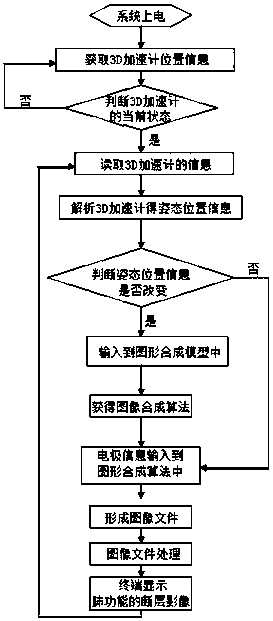
[0016] See figure 1 , the electrical impedance tomography lung imaging method based on the 3D accelerometer, firstly, after the electrical impedance tomography lung imaging system is powered on, obtain the position information of the 3D accelerometer, and adjust the position of the 3D accelerometer on the non-contact electrical impedance sensor until it is determined to be suitable The installation position is fixed. The non-contact electrical impedance sensor...


PUM


Abstract

The invention discloses an EIT (electrical impedance tomography) lung imaging method based on a 3D accelerometer. The method comprises steps as follows: posture position information of a to-be-detected person is collected by the 3D accelerometer in real time and is transmitted to a controller of an EIT lung imaging system together with current electrode information; the controller judges whether the position information is changed according to the posture position information, and when the position information is changed, new position information is input into an image synthesis model, so that a new image synthesis algorithm is obtained; the currently collected electrode information is input into the new image synthesis algorithm, an image file is formed, finally, the image file is processed, and a tomographic image of the lung function is obtained and displayed by a terminal. The 3D accelerometer is adopted to collect the posture position information in real time, so that a doctor is not required to input related posture information manually and frequently; besides, the calculating accuracy of the 3D accelerometer is high, accordingly, continuity of an intensive care process is guaranteed, and the problem of inaccuracy caused by the fact a patient changes the posture himself in a long-term intensive care process is solved.

Description

technical field [0001] The invention relates to the field of electrical impedance tomography lung imaging system, in particular to a 3D accelerometer-based electrical impedance tomography lung imaging method. Background technique [0002] Bioelectrical impedance imaging technology uses the electrical characteristics and changes of biological tissues or organs to extract biomedical information related to pathological conditions. It is a new generation of functional imaging technology that has emerged in the past 20 years after morphology and structure imaging. , continuous, no radiation, fast response, low cost and so on. The principle of bioelectrical impedance tomography lung imaging system is to inject weak alternating current into the human body, and the current will flow along the path of least resistance, thus forming a potential on the surface of the body, using electrodes to continuously measure the potential, and then converting it into the lung function tomography....

Claims


Application Information

Patent Timeline
Patent Type & Authority Patents(China)
IPC IPC(8): A61B5/11
CPCA61B5/0033A61B5/0536A61B5/1114A61B5/1116A61B5/6805A61B2576/02
Inventor 丁天赦方建新
Owner 合肥诺和电子科技有限公司
Why Eureka
Try Eureka