Hysteroscope and biopsy forceps integrated sleeve device capable of preventing falling during biopsy

A cannula device and biopsy forceps technology, applied in endoscope, colposcopy, medical science and other directions, can solve the problems of increased operation difficulty and time, insufficient clamping force, double protection, etc., to avoid repeated surgical operations, Increase the success rate and ensure the anti-slip effect

Inactive Publication Date: 2020-09-15
THE FIRST AFFILIATED HOSPITAL OF ZHENGZHOU UNIV
View PDF0 Cites 6 Cited by
  • Summary
  • Abstract
  • Description
  • Claims
  • Application Information

AI Technical Summary

Problems solved by technology

Most biopsy forceps are limited to a single sample due to increased surgical difficulty and time due to the limited space between the two jaws of the biopsy forceps
[0004] 2. When the existing biopsy forceps are in the process of grasping the biopsy inside the uterine cavity, when encountering larger tissues, it often occurs that the biopsy forceps are too small or the clamping force is insufficient to cause the biopsy tissue to fall from the biopsy forceps during the removal process. Slippage, leading to tissue sampling failure, often requires doctors to re-access the biopsy forceps device to clamp the diseased biopsy tissue, which greatly affects the efficiency of the operation, prolongs unnecessary operation time, and is also a pain for the patient
[0005] 3. At present, the jaws of biopsy forceps on the market are designed with different serrations or jaw shapes, which are used for different biopsy situations, but there is no double protection for the biopsy forceps to prevent falling off, and the biopsy tissue may fall off. re-access gripping

Method used


Image

  • Hysteroscope and biopsy forceps integrated sleeve device capable of preventing falling during biopsy
  • Hysteroscope and biopsy forceps integrated sleeve device capable of preventing falling during biopsy
  • Hysteroscope and biopsy forceps integrated sleeve device capable of preventing falling during biopsy

Examples


Embodiment Construction

[0041]In order to enable those skilled in the art to better understand the solutions of the present invention, the following will clearly and completely describe the technical solutions in the embodiments of the present invention in conjunction with the drawings in the embodiments of the present invention. Obviously, the described embodiments are only It is an embodiment of a part of the present invention, but not all embodiments. Based on the embodiments of the present invention, all other embodiments obtained by persons of ordinary skill in the art without making creative efforts shall fall within the protection scope of the present invention.

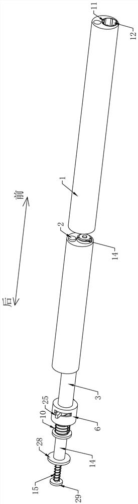
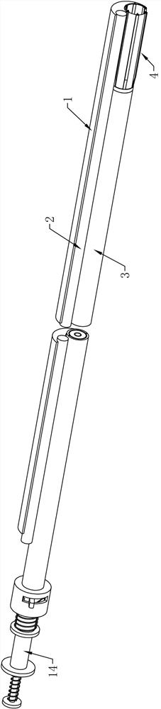
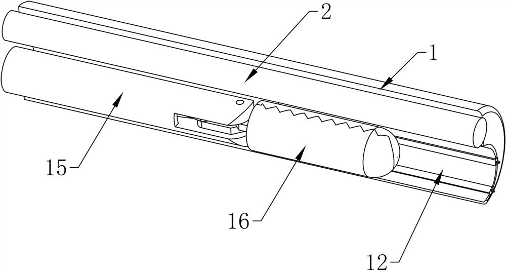
[0042] Such as Figures 1 to 17 As shown, a hysteroscopic biopsy forceps integrated cannula device for preventing biopsy from falling off according to an embodiment of the present invention includes: cannula I1, hysteroscope 2, cannula II3, claw sleeve 4, wire drawing ring 5, wire drawing sleeve Tube, wire drawing, casing 6, guide t...


PUM


Abstract

The invention relates to the field of medical instruments, in particular to a hysteroscope and biopsy forceps integrated sleeve device capable of preventing falling during biopsy. The hysteroscope andbiopsy forceps integrated sleeve device comprises a sleeve I, a hysteroscope, a sleeve II, a claw sleeve, a wire drawing ring, wire drawing sleeves, drawing wires, a sleeve shell, a guide pipe, a ratchet top plate I, a ratchet top plate II, a spring I and a biopsy forceps structure. Three states of the claw sleeve in the device respectively correspond to three stages of biopsy grabbing of biopsyforceps in a targeted manner. In the folding state I, the biopsy forceps are contracted into the sleeve I before grabbing. In the state II, due to the elastic opening characteristic of the claw sleeve, the claw sleeve can be automatically opened when stretching out, biopsy tissue grabbed by the biopsy forceps can be covered with the claw sleeve, and after elastic opening, biopsy tissue with different sizes can be covered in a universal manner through the open claw sleeve. In the state III, a port at the front end of the claw sleeve is pulled back by the drawing wires to be tightened and closedinto a closed state, so that the clamped biopsy tissue is completely wrapped, and double guarantees are provided for preventing the biopsy tissue from falling off.

Description

technical field [0001] The invention relates to the field of medical devices, in particular to a hysteroscopic biopsy forceps integrated sleeve device capable of preventing biopsy from falling off. Background technique [0002] Hysteroscopy is a new minimally invasive gynecological diagnosis and treatment technology. It is a kind of fiber light source endoscope used for inspection and treatment of uterine cavity, including hysteroscope, energy system, light source system, perfusion system and imaging system. It uses the front part of the mirror body to enter the uterine cavity, and has a magnifying effect on the observed parts. It is intuitive and accurate and has become the preferred inspection method for gynecological hemorrhagic diseases and intrauterine lesions. Through hysteroscopy, a series of detection and treatment such as examination, diagnosis and analysis are carried out on women. Hysteroscopy and surgery are the best options for determining whether there are les...

Claims


Application Information

Patent Timeline
Patent Type & Authority Applications(China)
IPC IPC(8): A61B1/303A61B10/04A61B10/06A61B1/04
CPCA61B1/303A61B1/00131A61B1/00064A61B10/04A61B10/06A61B10/0291A61B1/04Y02A40/81
Inventor 方兰兰孙莹璞郑融键
Owner THE FIRST AFFILIATED HOSPITAL OF ZHENGZHOU UNIV
Why Eureka
Try Eureka